 Movie
Movie Controller

[English] 日本語
 Yorodumi
- PDB-1nf8: Crystal structure of PhzD protein active site mutant with substrate -
+ Open data

- Basic information
| Entry | Database: PDB / ID: 1nf8 | ||||||
|---|---|---|---|---|---|---|---|
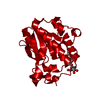
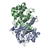
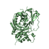
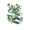
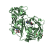
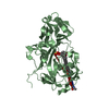
| Title | Crystal structure of PhzD protein active site mutant with substrate | ||||||
 Components Components | phenazine biosynthesis protein phzD | ||||||
 Keywords Keywords | HYDROLASE / isochorismatase / enzyme / phenazine pathway | ||||||
| Function / homology |  Function and homology information Function and homology informationtrans-2,3-dihydro-3-hydroxyanthranilic acid synthase / isochorismatase activity / phenazine biosynthetic process / cytosol Similarity search - Function | ||||||
| Biological species |   Pseudomonas aeruginosa (bacteria) Pseudomonas aeruginosa (bacteria) | ||||||
| Method |  X-RAY DIFFRACTION / MOLECULAR REPLACEMENT / Resolution: 1.6 Å X-RAY DIFFRACTION / MOLECULAR REPLACEMENT / Resolution: 1.6 Å | ||||||
 Authors Authors | Parsons, F. / Calabrese, K. / Eisenstein, E. / Ladner, J.E. | ||||||
 Citation Citation | Journal: Biochemistry / Year: 2003 Title: Structure and mechanism of Pseudomonas aeruginosa PhzD, an isochorismatase from the phenazine biosynthetic pathway Authors: Parsons, J.F. / Calabrese, K. / Eisenstein, E. / Ladner, J.E. | ||||||
| History |
|
- Structure visualization
| Structure viewer | Molecule: MolmilJmol/JSmol |
|---|
- Downloads & links
-Download
| PDBx/mmCIF format | 1nf8.cif.gz | 109.8 KB | Display | PDBx/mmCIF format |
|---|---|---|---|---|
| PDB format | pdb1nf8.ent.gz | 84 KB | Display | PDB format |
| PDBx/mmJSON format | 1nf8.json.gz | Tree view | PDBx/mmJSON format | |
| Others |  Other downloads Other downloads |
-Validation report
| Arichive directory | https://data.pdbj.org/pub/pdb/validation_reports/nf/1nf8ftp://data.pdbj.org/pub/pdb/validation_reports/nf/1nf8 | HTTPS FTP |
|---|
-Related structure data


-Links
 PDBj
PDBj- Assembly
| Deposited unit | 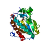
| |||||||||
|---|---|---|---|---|---|---|---|---|---|---|
| 1 | 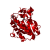
| |||||||||
| Unit cell |
| |||||||||
| Components on special symmetry positions |
| |||||||||
| Details | The second part of the biological assembly is generated by the two fold axis: -x,y,-z+3/2 |
-Components
| #1: Protein | Mass: 23176.529 Da / Num. of mol.: 1 / Mutation: D38A Source method: isolated from a genetically manipulated source Source: (gene. exp.) Pseudomonas aeruginosa (bacteria) / Gene: PhzD / Plasmid: pET28a / Species (production host): Escherichia coli / Production host: References: UniProt: Q7DC80, UniProt: P0DPB9*PLUS, isochorismatase |
|---|---|
| #2: Sugar | ChemComp-BOG /   Type: D-saccharide / Mass: 292.369 Da / Num. of mol.: 1 Type: D-saccharide / Mass: 292.369 Da / Num. of mol.: 1Source method: isolated from a genetically manipulated source Formula: C14H28O6 / Comment: detergent*YM |
| #3: Chemical | ChemComp-ISC / (  Mass: 226.183 Da / Num. of mol.: 1 / Source method: obtained synthetically / Formula: C10H10O6 Mass: 226.183 Da / Num. of mol.: 1 / Source method: obtained synthetically / Formula: C10H10O6 |
| #4: Water | ChemComp-HOH /  Mass: 18.015 Da / Num. of mol.: 298 / Source method: isolated from a natural source / Formula: H2O Mass: 18.015 Da / Num. of mol.: 298 / Source method: isolated from a natural source / Formula: H2O |
-Experimental details
-Experiment
| Experiment | Method: X-RAY DIFFRACTION / Number of used crystals: 1 |
|---|
- Sample preparation
| Crystal | Density Matthews: 2.02 Å3/Da / Density % sol: 47.45 % | ||||||||||||||||||||||||||||||
|---|---|---|---|---|---|---|---|---|---|---|---|---|---|---|---|---|---|---|---|---|---|---|---|---|---|---|---|---|---|---|---|
| Crystal grow | Temperature: 298 K / Method: vapor diffusion, hanging drop / pH: 7 Details: 10-20% polyethylene glycol 4000, 0.2M ammonium formate, 0.2% beta-octylglucoside, 1mM isochorismate, pH 7, VAPOR DIFFUSION, HANGING DROP, temperature 298K | ||||||||||||||||||||||||||||||
| Crystal grow | *PLUS Method: vapor diffusion, hanging drop | ||||||||||||||||||||||||||||||
| Components of the solutions | *PLUS
|
-Data collection
| Diffraction | Mean temperature: 100 K |
|---|---|
| Diffraction source | Source: ROTATING ANODE / Type: RIGAKU MICROMAX-007 / Wavelength: 1.5418 Å |
| Detector | Type: RIGAKU RAXIS IV++ / Detector: IMAGE PLATE / Date: Jul 15, 2002 / Details: OSMIC |
| Radiation | Monochromator: OSMIC / Protocol: SINGLE WAVELENGTH / Monochromatic (M) / Laue (L): M / Scattering type: x-ray |
| Radiation wavelength | Wavelength: 1.5418 Å / Relative weight: 1 |
| Reflection | Resolution: 1.6→20 Å / Num. obs: 27572 / % possible obs: 99.7 % / Redundancy: 3 % / Rmerge(I) obs: 0.042 / Net I/σ(I): 22 |
| Reflection shell | Resolution: 1.6→1.7 Å / Redundancy: 3 % / Rmerge(I) obs: 0.207 / Mean I/σ(I) obs: 3.1 / Num. unique all: 3895 / % possible all: 99 |
| Reflection | *PLUS Num. measured all: 82842 |
| Reflection shell | *PLUS % possible obs: 99 % |
- Processing
| Software |
| |||||||||||||||||||||||||||||||||
|---|---|---|---|---|---|---|---|---|---|---|---|---|---|---|---|---|---|---|---|---|---|---|---|---|---|---|---|---|---|---|---|---|---|---|
| Refinement | Method to determine structure: MOLECULAR REPLACEMENT Starting model: Native structure Resolution: 1.6→20 Å / Num. parameters: 17662 / Num. restraintsaints: 21130 / Cross valid method: FREE R / σ(F): 0 / Stereochemistry target values: ENGH AND HUBER
| |||||||||||||||||||||||||||||||||
| Refine analyze | Num. disordered residues: 0 / Occupancy sum hydrogen: 1608 / Occupancy sum non hydrogen: 1962 | |||||||||||||||||||||||||||||||||
| Refinement step | Cycle: LAST / Resolution: 1.6→20 Å
| |||||||||||||||||||||||||||||||||
| Refine LS restraints |
| |||||||||||||||||||||||||||||||||
| Software | *PLUS Name: SHELXL / Version: 97 / Classification: refinement | |||||||||||||||||||||||||||||||||
| Refinement | *PLUS Highest resolution: 1.6 Å / Lowest resolution: 20 Å / % reflection Rfree: 5 % / Rfactor Rfree: 0.21 | |||||||||||||||||||||||||||||||||
| Solvent computation | *PLUS | |||||||||||||||||||||||||||||||||
| Displacement parameters | *PLUS | |||||||||||||||||||||||||||||||||
| Refine LS restraints | *PLUS
|
