 Movie
Movie Controller

[English] 日本語
 Yorodumi
- PDB-1oa2: Comparison of Family 12 Glycoside Hydrolases and Recruited Substi... -
+ Open data

- Basic information
| Entry | Database: PDB / ID: 1oa2 | |||||||||
|---|---|---|---|---|---|---|---|---|---|---|
| Title | Comparison of Family 12 Glycoside Hydrolases and Recruited Substitutions Important for Thermal Stability | |||||||||
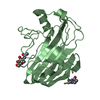
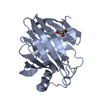
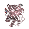
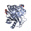
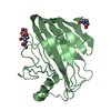
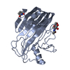
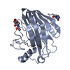
 Components Components | ENDO-BETA-1,4-GLUCANASE | |||||||||
 Keywords Keywords | HYDROLASE / CELLULASE / CELLULOSE DEGRADATION / ENDOGLUCANASE / GLYCOSYL HYDROLASE / GH FAMILY 12 / TRICHODERMA REESEI CEL12A | |||||||||
| Function / homology |  Function and homology information Function and homology information | |||||||||
| Biological species |  TRICHODERMA REESEI (fungus) TRICHODERMA REESEI (fungus) | |||||||||
| Method |  X-RAY DIFFRACTION / SYNCHROTRON / MOLECULAR REPLACEMENT / Resolution: 1.5 Å X-RAY DIFFRACTION / SYNCHROTRON / MOLECULAR REPLACEMENT / Resolution: 1.5 Å | |||||||||
 Authors Authors | Sandgren, M. / Gualfetti, P.J. / Shaw, A. / Gross, L.S. / Saldajeno, M. / Day, A.G. / Jones, T.A. / Mitchinson, C. | |||||||||
 Citation Citation | Journal: Protein Sci. / Year: 2003 Title: Comparison of Family 12 Glycoside Hydrolases and Recruited Substitutions Important for Thermal Stability Authors: Sandgren, M. / Gualfetti, P.J. / Shaw, A. / Gross, L.S. / Saldajeno, M. / Day, A.G. / Jones, T.A. / Mitchinson, C. | |||||||||
| History |
| |||||||||
| Remark 700 | SHEET THE SHEET STRUCTURE OF THIS MOLECULE IS BIFURCATED. IN ORDER TO REPRESENT THIS FEATURE IN ... SHEET THE SHEET STRUCTURE OF THIS MOLECULE IS BIFURCATED. IN ORDER TO REPRESENT THIS FEATURE IN THE SHEET RECORDS BELOW, TWO SHEETS ARE DEFINED. |
- Structure visualization
| Structure viewer | Molecule: MolmilJmol/JSmol |
|---|
- Downloads & links
-Download
| PDBx/mmCIF format | 1oa2.cif.gz | 260.8 KB | Display | PDBx/mmCIF format |
|---|---|---|---|---|
| PDB format | pdb1oa2.ent.gz | 214.1 KB | Display | PDB format |
| PDBx/mmJSON format | 1oa2.json.gz | Tree view | PDBx/mmJSON format | |
| Others |  Other downloads Other downloads |
-Validation report
| Arichive directory | https://data.pdbj.org/pub/pdb/validation_reports/oa/1oa2ftp://data.pdbj.org/pub/pdb/validation_reports/oa/1oa2 | HTTPS FTP |
|---|
-Related structure data
| Related structure data |  1oa3C  1oa4C  1h8vS S: Starting model for refinement C: citing same article ( |
|---|---|
| Similar structure data |


-Links
 PDBj
PDBj
- Assembly
| Deposited unit | 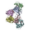
| ||||||||
|---|---|---|---|---|---|---|---|---|---|
| 1 | 
| ||||||||
| 2 | 
| ||||||||
| 3 | 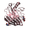
| ||||||||
| 4 | 
| ||||||||
| 5 | 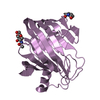
| ||||||||
| 6 | 
| ||||||||
| Unit cell |
|
-Components
| #1: Protein | Mass: 23501.395 Da / Num. of mol.: 6 / Fragment: RESIDUES 17-234 / Mutation: YES Source method: isolated from a genetically manipulated source Source: (gene. exp.) TRICHODERMA REESEI (fungus) / Production host: #2: Sugar | ChemComp-NAG /   Type: D-saccharide, beta linking / Mass: 221.208 Da / Num. of mol.: 6 Type: D-saccharide, beta linking / Mass: 221.208 Da / Num. of mol.: 6Source method: isolated from a genetically manipulated source Formula: C8H15NO6 #3: Water | ChemComp-HOH / |  Mass: 18.015 Da / Num. of mol.: 641 / Source method: isolated from a natural source / Formula: H2O Mass: 18.015 Da / Num. of mol.: 641 / Source method: isolated from a natural source / Formula: H2OCompound details | ENDOHYDROL | Has protein modification | Y | Sequence details | GLN 17 MODIFIED RESIDUE | |
|---|
-Experimental details
-Experiment
| Experiment | Method: X-RAY DIFFRACTION / Number of used crystals: 1 |
|---|
- Sample preparation
| Crystal | Density Matthews: 2 Å3/Da / Density % sol: 40 % | ||||||||||||||||||||||||||||||
|---|---|---|---|---|---|---|---|---|---|---|---|---|---|---|---|---|---|---|---|---|---|---|---|---|---|---|---|---|---|---|---|
| Crystal grow | pH: 6 / Details: pH 6.00 | ||||||||||||||||||||||||||||||
| Crystal grow | *PLUS Temperature: 20-24 ℃ / Method: vapor diffusion, hanging drop | ||||||||||||||||||||||||||||||
| Components of the solutions | *PLUS
|
-Data collection
| Diffraction | Mean temperature: 100 K |
|---|---|
| Diffraction source | Source: SYNCHROTRON / Site: ESRF  / Beamline: ID14-1 / Wavelength: 0.93 / Beamline: ID14-1 / Wavelength: 0.93 |
| Detector | Type: MARRESEARCH / Detector: CCD / Date: Dec 3, 2000 |
| Radiation | Protocol: SINGLE WAVELENGTH / Monochromatic (M) / Laue (L): M / Scattering type: x-ray |
| Radiation wavelength | Wavelength: 0.93 Å / Relative weight: 1 |
| Reflection | Resolution: 1.5→25 Å / Num. obs: 172895 / % possible obs: 94 % / Observed criterion σ(I): 3 / Redundancy: 2.8 % / Rsym value: 0.037 / Net I/σ(I): 24.9 |
| Reflection shell | Resolution: 1.5→1.53 Å / Mean I/σ(I) obs: 3 / Rsym value: 0.38 / % possible all: 93 |
| Reflection | *PLUS Highest resolution: 1.5 Å / Lowest resolution: 25 Å / % possible obs: 94.4 % / Num. measured all: 490560 / Rmerge(I) obs: 0.037 |
| Reflection shell | *PLUS % possible obs: 94.3 % / Rmerge(I) obs: 0.381 / Mean I/σ(I) obs: 2.3 |
- Processing
| Software |
| ||||||||||||||||||||
|---|---|---|---|---|---|---|---|---|---|---|---|---|---|---|---|---|---|---|---|---|---|
| Refinement | Method to determine structure: MOLECULAR REPLACEMENT Starting model: PDB ENTRY 1H8V Resolution: 1.5→20 Å / SU B: 2.56872 / SU ML: 0.09565 / Cross valid method: THROUGHOUT / σ(F): 0 / ESU R: 0.15274 / ESU R Free: 0.12287 / Details: HYDROGENS HAVE BEEN ADDED IN THE RIDING POSITIONS
| ||||||||||||||||||||
| Displacement parameters | Biso mean: 17.86 Å2
| ||||||||||||||||||||
| Refinement step | Cycle: LAST / Resolution: 1.5→20 Å
| ||||||||||||||||||||
| Refinement | *PLUS Rfactor Rfree: 0.219 / Rfactor Rwork: 0.201 | ||||||||||||||||||||
| Solvent computation | *PLUS | ||||||||||||||||||||
| Displacement parameters | *PLUS | ||||||||||||||||||||
| Refine LS restraints | *PLUS
|
