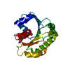
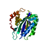
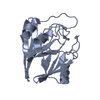
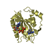
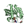
- PDB-2nla: Crystal structure of the Mcl-1:mNoxaB BH3 complex -
+
Open data
ID or keywords:
Loading...
-
Basic information
Entry
Database: PDB / ID: 2nla
Title
Crystal structure of the Mcl-1:mNoxaB BH3 complex
Components
FUSION PROTEIN CONSISTING OF Induced myeloid leukemia cell differentiation protein Mcl-1 homolog
Phorbol-12-myristate-13-acetate-induced protein 1
Keywords
APOPTOSIS / Bcl-2 / Mcl-1 / Noxa
Function / homology
Function and homology information
Activation of NOXA and translocation to mitochondria / BH3-only proteins associate with and inactivate anti-apoptotic BCL-2 members / response to dsRNA / positive regulation of extrinsic apoptotic signaling pathway via death domain receptors / BH domain binding / positive regulation of protein localization to mitochondrion / positive regulation of glucose metabolic process / positive regulation of oxidative stress-induced neuron intrinsic apoptotic signaling pathway / cell fate determination / cellular homeostasis ...Activation of NOXA and translocation to mitochondria / BH3-only proteins associate with and inactivate anti-apoptotic BCL-2 members / response to dsRNA / positive regulation of extrinsic apoptotic signaling pathway via death domain receptors / BH domain binding / positive regulation of protein localization to mitochondrion / positive regulation of glucose metabolic process / positive regulation of oxidative stress-induced neuron intrinsic apoptotic signaling pathway / cell fate determination / cellular homeostasis / regulation of mitochondrial membrane permeability / mitochondrial fusion / Bcl-2 family protein complex / positive regulation of DNA damage response, signal transduction by p53 class mediator / T cell homeostasis / positive regulation of release of cytochrome c from mitochondria / apoptotic mitochondrial changes / negative regulation of mitochondrial membrane potential / intrinsic apoptotic signaling pathway by p53 class mediator / negative regulation of anoikis / BH3 domain binding / intrinsic apoptotic signaling pathway in response to endoplasmic reticulum stress / transmembrane protein transporter activity / intrinsic apoptotic signaling pathway in response to DNA damage by p53 class mediator / negative regulation of extrinsic apoptotic signaling pathway in absence of ligand / response to X-ray / response to UV / negative regulation of fibroblast proliferation / cellular response to glucose starvation / extrinsic apoptotic signaling pathway in absence of ligand / positive regulation of intrinsic apoptotic signaling pathway / proteasomal protein catabolic process / response to cytokine / reactive oxygen species metabolic process / intrinsic apoptotic signaling pathway / release of cytochrome c from mitochondria / negative regulation of autophagy / intrinsic apoptotic signaling pathway in response to DNA damage / Signaling by ALK fusions and activated point mutants / positive regulation of neuron apoptotic process / channel activity / Interleukin-4 and Interleukin-13 signaling / regulation of apoptotic process / defense response to virus / cell differentiation / mitochondrial outer membrane / protein dimerization activity / positive regulation of apoptotic process / protein heterodimerization activity / apoptotic process / DNA damage response / negative regulation of apoptotic process / protein-containing complex binding / mitochondrion / nucleoplasm / membrane / nucleus / cytoplasm / cytosol Similarity search - Function
Phorbol-12-myristate-13-acetate-induced protein 1 / Phorbol-12-myristate-13-acetate-induced / Apoptosis regulator, Mcl-1 / Blc2-like / Apoptosis Regulator Bcl-x / Apoptosis regulator, Bcl-2, BH3 motif, conserved site / Apoptosis regulator, Bcl-2 family BH3 motif signature. / Apoptosis regulator, Bcl-2, BH1 motif, conserved site / Apoptosis regulator, Bcl-2 family BH1 motif signature. / Apoptosis regulator, Bcl-2, BH2 motif, conserved site ...Phorbol-12-myristate-13-acetate-induced protein 1 / Phorbol-12-myristate-13-acetate-induced / Apoptosis regulator, Mcl-1 / Blc2-like / Apoptosis Regulator Bcl-x / Apoptosis regulator, Bcl-2, BH3 motif, conserved site / Apoptosis regulator, Bcl-2 family BH3 motif signature. / Apoptosis regulator, Bcl-2, BH1 motif, conserved site / Apoptosis regulator, Bcl-2 family BH1 motif signature. / Apoptosis regulator, Bcl-2, BH2 motif, conserved site / Apoptosis regulator, Bcl-2 family BH2 motif signature. / Bcl-2 family / BCL (B-Cell lymphoma); contains BH1, BH2 regions / Bcl2-like / Bcl-2, Bcl-2 homology region 1-3 / Apoptosis regulator proteins, Bcl-2 family / BCL2-like apoptosis inhibitors family profile. / Bcl-2-like superfamily / Orthogonal Bundle / Mainly Alpha Similarity search - Domain/homology
Induced myeloid leukemia cell differentiation protein Mcl-1 homolog / Induced myeloid leukemia cell differentiation protein Mcl-1 / Phorbol-12-myristate-13-acetate-induced protein 1 Similarity search - Component
A: FUSION PROTEIN CONSISTING OF Induced myeloid leukemia cell differentiation protein Mcl-1 homolog B: Phorbol-12-myristate-13-acetate-induced protein 1
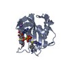
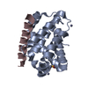
Method to determine structure: MOLECULAR REPLACEMENT / Resolution: 2.8→73.92 Å / Cor.coef. Fo:Fc: 0.955 / Cor.coef. Fo:Fc free: 0.873 / SU B: 55.937 / SU ML: 0.472 / Cross valid method: THROUGHOUT / σ(F): 0 / ESU R Free: 0.452 / Stereochemistry target values: MAXIMUM LIKELIHOOD Details: CHAIN D REFERS TO THE NOXA PEPTIDE WHICH IS COVALENTLY LINKED TO MCL-1 THROUGH CYS 286 OF MCL-1 AND CYS 75 OF NOXA. HYDROGENS HAVE BEEN ADDED IN THE RIDING POSITIONS.
Rfactor
Num. reflection
% reflection
Selection details
Rfree
0.291
230
4.7 %
RANDOM
Rwork
0.207
-
-
-
obs
0.211
4699
99.86 %
-
Solvent computation
Ion probe radii: 0.8 Å / Shrinkage radii: 0.8 Å / VDW probe radii: 1.2 Å / Solvent model: MASK
Displacement parameters
Biso mean: 80.22 Å2
Baniso -1
Baniso -2
Baniso -3
1-
1.36 Å2
0.68 Å2
0 Å2
2-
-
1.36 Å2
0 Å2
3-
-
-
-2.04 Å2
Refinement step
Cycle: LAST / Resolution: 2.8→73.92 Å
Protein
Nucleic acid
Ligand
Solvent
Total
Num. atoms
1375
0
0
31
1406
Refine LS restraints
Refine-ID
Type
Dev ideal
Dev ideal target
Number
X-RAY DIFFRACTION
r_bond_refined_d
0.012
0.022
1395
X-RAY DIFFRACTION
r_angle_refined_deg
1.341
1.951
1874
X-RAY DIFFRACTION
r_dihedral_angle_1_deg
5.571
5
169
X-RAY DIFFRACTION
r_dihedral_angle_2_deg
34.213
22.958
71
X-RAY DIFFRACTION
r_dihedral_angle_3_deg
19.407
15
264
X-RAY DIFFRACTION
r_dihedral_angle_4_deg
16.516
15
17
X-RAY DIFFRACTION
r_chiral_restr
0.088
0.2
208
X-RAY DIFFRACTION
r_gen_planes_refined
0.004
0.02
1044
X-RAY DIFFRACTION
r_nbd_refined
0.242
0.2
717
X-RAY DIFFRACTION
r_nbtor_refined
0.303
0.2
947
X-RAY DIFFRACTION
r_xyhbond_nbd_refined
0.204
0.2
68
X-RAY DIFFRACTION
r_symmetry_vdw_refined
0.208
0.2
47
X-RAY DIFFRACTION
r_symmetry_hbond_refined
0.226
0.2
9
X-RAY DIFFRACTION
r_mcbond_it
0.425
1.5
875
X-RAY DIFFRACTION
r_mcangle_it
0.705
2
1353
X-RAY DIFFRACTION
r_scbond_it
0.949
3
590
X-RAY DIFFRACTION
r_scangle_it
1.561
4.5
521
LS refinement shell
Resolution: 2.798→2.871 Å / Total num. of bins used: 20
Rfactor
Num. reflection
% reflection
Rfree
0.428
18
-
Rwork
0.413
344
-
obs
-
362
98.91 %
Refinement TLS params.
Method: refined / Refine-ID: X-RAY DIFFRACTION
ID
L11 (°2)
L12 (°2)
L13 (°2)
L22 (°2)
L23 (°2)
L33 (°2)
S11 (Å °)
S12 (Å °)
S13 (Å °)
S21 (Å °)
S22 (Å °)
S23 (Å °)
S31 (Å °)
S32 (Å °)
S33 (Å °)
T11 (Å2)
T12 (Å2)
T13 (Å2)
T22 (Å2)
T23 (Å2)
T33 (Å2)
Origin x (Å)
Origin y (Å)
Origin z (Å)
1
7.3458
-0.5017
-0.6155
5.7216
0.085
10.5334
-0.0137
-0.4616
0.776
-0.1552
0.1268
-0.0328
-0.9296
-0.6959
-0.1132
-0.3586
0.061
0.031
-0.3017
-0.0586
-0.323
-26.6062
3.2318
-7.1963
2
20.4622
-9.6652
3.5592
20.4276
0.3025
7.7655
0.5758
1.2816
-0.6782
-1.8879
-0.0637
1.5006
-0.1188
0.0764
-0.5121
-0.1677
-0.0108
0.0187
-0.1562
-0.1316
-0.4211
-18.7253
20.7181
4.2364
Refinement TLS group
Refine-ID: X-RAY DIFFRACTION / Selection: ALL
ID
Refine TLS-ID
Auth asym-ID
Label asym-ID
Auth seq-ID
Label seq-ID
1
1
A
A
175 - 321
5 - 151
2
2
B
B
73 - 93
6 - 26
+
About Yorodumi
-
News
-
Feb 9, 2022. New format data for meta-information of EMDB entries
New format data for meta-information of EMDB entries
Version 3 of the EMDB header file is now the official format.
The previous official version 1.9 will be removed from the archive.
In the structure databanks used in Yorodumi, some data are registered as the other names, "COVID-19 virus" and "2019-nCoV". Here are the details of the virus and the list of structure data.
Jan 31, 2019. EMDB accession codes are about to change! (news from PDBe EMDB page)
EMDB accession codes are about to change! (news from PDBe EMDB page)
The allocation of 4 digits for EMDB accession codes will soon come to an end. Whilst these codes will remain in use, new EMDB accession codes will include an additional digit and will expand incrementally as the available range of codes is exhausted. The current 4-digit format prefixed with “EMD-” (i.e. EMD-XXXX) will advance to a 5-digit format (i.e. EMD-XXXXX), and so on. It is currently estimated that the 4-digit codes will be depleted around Spring 2019, at which point the 5-digit format will come into force.
The EM Navigator/Yorodumi systems omit the EMD- prefix.
Related info.:Q: What is EMD? / ID/Accession-code notation in Yorodumi/EM Navigator
Yorodumi is a browser for structure data from EMDB, PDB, SASBDB, etc.
This page is also the successor to EM Navigator detail page, and also detail information page/front-end page for Omokage search.
The word "yorodu" (or yorozu) is an old Japanese word meaning "ten thousand". "mi" (miru) is to see.
Related info.:EMDB / PDB / SASBDB / Comparison of 3 databanks / Yorodumi Search / Aug 31, 2016. New EM Navigator & Yorodumi / Yorodumi Papers / Jmol/JSmol / Function and homology information / Changes in new EM Navigator and Yorodumi
 Movie
Movie Controller


 Open data
 Basic information
 Components
 Keywords
 Function and homology information
 Homo sapiens (human)
 X-RAY DIFFRACTION /
  Authors
 Citation
 Structure visualization
 Downloads & links
 Other downloads


 PDBj
PDBj
 Assembly

 Mass: 18.015 Da / Num. of mol.: 31 / Source method: isolated from a natural source / Formula: H2O
 Sample preparation
 / Beamline: X29A / Wavelength: 0.9715 Å
 Processing