 Movie
Movie Controller

[English] 日本語
 Yorodumi
- PDB-5xch: Crystal structure of Wild type Vps29 complexed with Zn+2 from Ent... -
+ Open data

- Basic information
| Entry | Database: PDB / ID: 5xch | ||||||
|---|---|---|---|---|---|---|---|
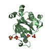
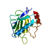
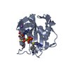
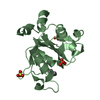
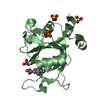
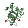
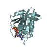
| Title | Crystal structure of Wild type Vps29 complexed with Zn+2 from Entamoeba histolytica | ||||||
 Components Components | Vacuolar protein sorting-associated protein 29 | ||||||
 Keywords Keywords | PROTEIN TRANSPORT / Entamoeba histolytica / Vacuolar protein sorting 29 / Metallophosphatase fold / Calcineurin-like phosphoesterase superfamily domain | ||||||
| Function / homology |  Function and homology information Function and homology informationretromer complex / retrograde transport, endosome to Golgi / protein transport / cytoplasmic vesicle / metal ion binding / cytosol Similarity search - Function | ||||||
| Biological species |   Entamoeba histolytica (eukaryote) Entamoeba histolytica (eukaryote) | ||||||
| Method |  X-RAY DIFFRACTION / SYNCHROTRON / MOLECULAR REPLACEMENT / Resolution: 2.85 Å X-RAY DIFFRACTION / SYNCHROTRON / MOLECULAR REPLACEMENT / Resolution: 2.85 Å | ||||||
 Authors Authors | Srivastava, V.K. / Yadav, R. / Tomar, P. / Gourinath, S. / Datta, S. | ||||||
 Citation Citation | Journal: Mol. Microbiol. / Year: 2017 Title: Structural and thermodynamic characterization of metal binding in Vps29 from Entamoeba histolytica: implication in retromer function. Authors: Srivastava, V.K. / Yadav, R. / Watanabe, N. / Tomar, P. / Mukherjee, M. / Gourinath, S. / Nakada-Tsukui, K. / Nozaki, T. / Datta, S. | ||||||
| History |
|
- Structure visualization
| Structure viewer | Molecule: MolmilJmol/JSmol |
|---|
- Downloads & links
-Download
| PDBx/mmCIF format | 5xch.cif.gz | 83.3 KB | Display | PDBx/mmCIF format |
|---|---|---|---|---|
| PDB format | pdb5xch.ent.gz | 61.3 KB | Display | PDB format |
| PDBx/mmJSON format | 5xch.json.gz | Tree view | PDBx/mmJSON format | |
| Others |  Other downloads Other downloads |
-Validation report
| Arichive directory | https://data.pdbj.org/pub/pdb/validation_reports/xc/5xchftp://data.pdbj.org/pub/pdb/validation_reports/xc/5xch | HTTPS FTP |
|---|
-Related structure data
| Related structure data | 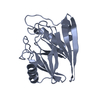 5xceSC 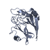 5xcjC 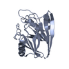 5xckC S: Starting model for refinement C: citing same article ( |
|---|---|
| Similar structure data |


-Links
 PDBj
PDBj
- Assembly
| Deposited unit | 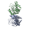
| ||||||||
|---|---|---|---|---|---|---|---|---|---|
| 1 | 
| ||||||||
| 2 | 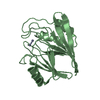
| ||||||||
| Unit cell |
|
-Components
| #1: Protein | Mass: 21876.969 Da / Num. of mol.: 2 Source method: isolated from a genetically manipulated source Details: HM-1:IMSS / Source: (gene. exp.) Entamoeba histolytica (eukaryote) / Gene: vsp, EhVPS29, CL6EHI_025270, EHI_025270 / Plasmid: pET28a / Production host:  #2: Chemical | ChemComp-ZN /   Mass: 65.409 Da / Num. of mol.: 4 / Source method: obtained synthetically / Formula: Zn Mass: 65.409 Da / Num. of mol.: 4 / Source method: obtained synthetically / Formula: Zn#3: Water | ChemComp-HOH / |  Mass: 18.015 Da / Num. of mol.: 25 / Source method: isolated from a natural source / Formula: H2O Mass: 18.015 Da / Num. of mol.: 25 / Source method: isolated from a natural source / Formula: H2O |
|---|
-Experimental details
-Experiment
| Experiment | Method: X-RAY DIFFRACTION / Number of used crystals: 1 |
|---|
- Sample preparation
| Crystal | Density Matthews: 2.19 Å3/Da / Density % sol: 43.72 % |
|---|---|
| Crystal grow | Temperature: 289 K / Method: vapor diffusion, hanging drop / pH: 6.5 Details: 10 % w/v PEG 20000, 20 % v/v PEGMME 550, 0.02 M of alcohol MIX, 0.1 M MES/Imidazole pH 6.5 |
-Data collection
| Diffraction | Mean temperature: 100 K |
|---|---|
| Diffraction source | Source: SYNCHROTRON / Site: ESRF  / Beamline: BM14 / Wavelength: 1.2 Å / Beamline: BM14 / Wavelength: 1.2 Å |
| Detector | Type: MAR CCD 165 mm / Detector: CCD / Date: Sep 23, 2016 |
| Radiation | Protocol: SINGLE WAVELENGTH / Monochromatic (M) / Laue (L): M / Scattering type: x-ray |
| Radiation wavelength | Wavelength: 1.2 Å / Relative weight: 1 |
| Reflection twin | Operator: h,-h-k,-l / Fraction: 0.45 |
| Reflection | Resolution: 2.85→40.9 Å / Num. obs: 8561 / % possible obs: 98 % / Redundancy: 11.2 % / Net I/σ(I): 21 |
- Processing
| Software |
| ||||||||||||||||||||||||||||
|---|---|---|---|---|---|---|---|---|---|---|---|---|---|---|---|---|---|---|---|---|---|---|---|---|---|---|---|---|---|
| Refinement | Method to determine structure: MOLECULAR REPLACEMENT Starting model: 5XCE Resolution: 2.85→40.9 Å / Cross valid method: FREE R-VALUE / σ(F): 2.06 / Phase error: 33.53
| ||||||||||||||||||||||||||||
| Solvent computation | Shrinkage radii: 0.9 Å / VDW probe radii: 1.11 Å | ||||||||||||||||||||||||||||
| Refinement step | Cycle: LAST / Resolution: 2.85→40.9 Å
| ||||||||||||||||||||||||||||
| Refine LS restraints |
| ||||||||||||||||||||||||||||
| LS refinement shell |
|
