 Movie
Movie Controller

[English] 日本語
 Yorodumi
- PDB-6ipb: Crystal Structure of the Cofactor-binding Domain of the Human Pha... -
+ Open data

- Basic information
| Entry | Database: PDB / ID: 6ipb | ||||||
|---|---|---|---|---|---|---|---|
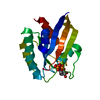
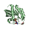
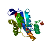
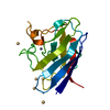
| Title | Crystal Structure of the Cofactor-binding Domain of the Human Phase II Drug Metabolism Enzyme UGT2B15 | ||||||
 Components Components | UDP-glucuronosyltransferase 2B15 | ||||||
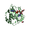
 Keywords Keywords | TRANSFERASE / Drug metabolism / UGT / Glycosyltransferase / Tartaric acid | ||||||
| Function / homology |  Function and homology information Function and homology informationglucuronosyltransferase / Glucuronidation / glucuronosyltransferase activity / Paracetamol ADME / estrogen metabolic process / Aspirin ADME / steroid metabolic process / xenobiotic metabolic process / endoplasmic reticulum membrane Similarity search - Function | ||||||
| Biological species |  Homo sapiens (human) Homo sapiens (human) | ||||||
| Method |  X-RAY DIFFRACTION / SYNCHROTRON / SAD / Resolution: 1.78 Å X-RAY DIFFRACTION / SYNCHROTRON / SAD / Resolution: 1.78 Å | ||||||
 Authors Authors | Zhang, L. / Xie, W. / Wang, C. | ||||||
 Citation Citation | Journal: Biochem. Pharmacol. / Year: 2019 Title: Insight into tartrate inhibition patterns in vitro and in vivo based on cocrystal structure with UDP-glucuronosyltransferase 2B15. Authors: Zhang, L. / Zhu, L. / Qu, W. / Wu, F. / Hu, M. / Xie, W. / Liu, Z. / Wang, C. | ||||||
| History |
|
- Structure visualization
| Structure viewer | Molecule: MolmilJmol/JSmol |
|---|
- Downloads & links
-Download
| PDBx/mmCIF format | 6ipb.cif.gz | 157.9 KB | Display | PDBx/mmCIF format |
|---|---|---|---|---|
| PDB format | pdb6ipb.ent.gz | 123.4 KB | Display | PDB format |
| PDBx/mmJSON format | 6ipb.json.gz | Tree view | PDBx/mmJSON format | |
| Others |  Other downloads Other downloads |
-Validation report
| Arichive directory | https://data.pdbj.org/pub/pdb/validation_reports/ip/6ipbftp://data.pdbj.org/pub/pdb/validation_reports/ip/6ipb | HTTPS FTP |
|---|
-Related structure data
| Similar structure data |
|---|


-Links
 PDBj
PDBj
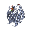
- Assembly
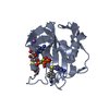
| Deposited unit | 
| ||||||||
|---|---|---|---|---|---|---|---|---|---|
| 1 | 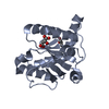
| ||||||||
| 2 | 
| ||||||||
| 3 | 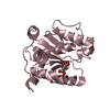
| ||||||||
| 4 | 
| ||||||||
| Unit cell |
|
-Components
| #1: Protein | Mass: 21342.635 Da / Num. of mol.: 4 Source method: isolated from a genetically manipulated source Source: (gene. exp.) Homo sapiens (human) / Gene: UGT2B15, UGT2B8 / Production host:  #2: Chemical | ChemComp-TLA /   Mass: 150.087 Da / Num. of mol.: 4 / Source method: obtained synthetically / Formula: C4H6O6 / Feature type: SUBJECT OF INVESTIGATION Mass: 150.087 Da / Num. of mol.: 4 / Source method: obtained synthetically / Formula: C4H6O6 / Feature type: SUBJECT OF INVESTIGATION#3: Chemical | ChemComp-GOL /   Mass: 92.094 Da / Num. of mol.: 4 / Source method: obtained synthetically / Formula: C3H8O3 / Feature type: SUBJECT OF INVESTIGATION Mass: 92.094 Da / Num. of mol.: 4 / Source method: obtained synthetically / Formula: C3H8O3 / Feature type: SUBJECT OF INVESTIGATION#4: Water | ChemComp-HOH / |  Mass: 18.015 Da / Num. of mol.: 655 / Source method: isolated from a natural source / Formula: H2O Mass: 18.015 Da / Num. of mol.: 655 / Source method: isolated from a natural source / Formula: H2O |
|---|
-Experimental details
-Experiment
| Experiment | Method: X-RAY DIFFRACTION / Number of used crystals: 1 |
|---|
- Sample preparation
| Crystal | Density Matthews: 2.13 Å3/Da / Density % sol: 42.36 % |
|---|---|
| Crystal grow | Temperature: 298 K / Method: vapor diffusion, sitting drop Details: 18% PEG 3350, 0.2M Na-Tartrate, 0.2M KCl, 2% glycerol |
-Data collection
| Diffraction | Mean temperature: 100 K / Serial crystal experiment: N |
|---|---|
| Diffraction source | Source: SYNCHROTRON / Site: SSRF  / Beamline: BL17U / Wavelength: 0.979 Å / Beamline: BL17U / Wavelength: 0.979 Å |
| Detector | Type: ADSC QUANTUM 315r / Detector: CCD / Date: May 6, 2018 |
| Radiation | Protocol: SINGLE WAVELENGTH / Monochromatic (M) / Laue (L): M / Scattering type: x-ray |
| Radiation wavelength | Wavelength: 0.979 Å / Relative weight: 1 |
| Reflection | Resolution: 1.775→70.91 Å / Num. obs: 64886 / % possible obs: 99.25 % / Redundancy: 6.6 % / CC1/2: 0.991 / Rpim(I) all: 0.04 / Rrim(I) all: 0.104 / Net I/σ(I): 22.64 |
| Reflection shell | Resolution: 1.78→1.84 Å / Mean I/σ(I) obs: 2.737 / Num. unique obs: 6779 / CC1/2: 0.902 / Rpim(I) all: 0.206 / Rrim(I) all: 0.521 |
- Processing
| Software |
| ||||||||||||||||||||||||||||||||||||||||||||||||||||||||||||||||||||||||||||||||||||||||||||||||||||||||||||||||||||||||||||||||||||||||||||||||||||||||||||||||||||||||||||||||||||||
|---|---|---|---|---|---|---|---|---|---|---|---|---|---|---|---|---|---|---|---|---|---|---|---|---|---|---|---|---|---|---|---|---|---|---|---|---|---|---|---|---|---|---|---|---|---|---|---|---|---|---|---|---|---|---|---|---|---|---|---|---|---|---|---|---|---|---|---|---|---|---|---|---|---|---|---|---|---|---|---|---|---|---|---|---|---|---|---|---|---|---|---|---|---|---|---|---|---|---|---|---|---|---|---|---|---|---|---|---|---|---|---|---|---|---|---|---|---|---|---|---|---|---|---|---|---|---|---|---|---|---|---|---|---|---|---|---|---|---|---|---|---|---|---|---|---|---|---|---|---|---|---|---|---|---|---|---|---|---|---|---|---|---|---|---|---|---|---|---|---|---|---|---|---|---|---|---|---|---|---|---|---|---|---|
| Refinement | Method to determine structure: SAD / Resolution: 1.78→70.91 Å / Cor.coef. Fo:Fc: 0.975 / Cor.coef. Fo:Fc free: 0.962 / SU B: 2.425 / SU ML: 0.074 / Cross valid method: THROUGHOUT / ESU R: 0.1 / ESU R Free: 0.098 / Details: HYDROGENS HAVE BEEN ADDED IN THE RIDING POSITIONS
| ||||||||||||||||||||||||||||||||||||||||||||||||||||||||||||||||||||||||||||||||||||||||||||||||||||||||||||||||||||||||||||||||||||||||||||||||||||||||||||||||||||||||||||||||||||||
| Solvent computation | Ion probe radii: 0.8 Å / Shrinkage radii: 0.8 Å / VDW probe radii: 1.2 Å | ||||||||||||||||||||||||||||||||||||||||||||||||||||||||||||||||||||||||||||||||||||||||||||||||||||||||||||||||||||||||||||||||||||||||||||||||||||||||||||||||||||||||||||||||||||||
| Displacement parameters | Biso mean: 26.014 Å2
| ||||||||||||||||||||||||||||||||||||||||||||||||||||||||||||||||||||||||||||||||||||||||||||||||||||||||||||||||||||||||||||||||||||||||||||||||||||||||||||||||||||||||||||||||||||||
| Refinement step | Cycle: 1 / Resolution: 1.78→70.91 Å
| ||||||||||||||||||||||||||||||||||||||||||||||||||||||||||||||||||||||||||||||||||||||||||||||||||||||||||||||||||||||||||||||||||||||||||||||||||||||||||||||||||||||||||||||||||||||
| Refine LS restraints |
|