 Movie
Movie Controller


+ Open data

- Basic information
| Entry | Database: PDB / ID: 6ukl | ||||||
|---|---|---|---|---|---|---|---|
| Title | Crystal Structure of a DiB2-split Protein | ||||||
 Components Components | (Outer membrane lipoprotein ...) x 2 | ||||||
 Keywords Keywords | FLUORESCENT PROTEIN / lipocalin / beta barrel / split protein / fluorogen activating protein | ||||||
| Function / homology |  Function and homology information Function and homology informationresponse to stress / cell outer membrane / DNA damage response / lipid binding Similarity search - Function | ||||||
| Biological species |  | ||||||
| Method |  X-RAY DIFFRACTION / SYNCHROTRON / MOLECULAR REPLACEMENT / molecular replacement / Resolution: 2.02 Å X-RAY DIFFRACTION / SYNCHROTRON / MOLECULAR REPLACEMENT / molecular replacement / Resolution: 2.02 Å | ||||||
 Authors Authors | Bozhanova, N.G. / Meiler, J. | ||||||
| Funding support |  United States, 1items United States, 1items
| ||||||
 Citation Citation | Journal: Sci Rep / Year: 2020 Title: DiB-splits: nature-guided design of a novel fluorescent labeling split system. Authors: Bozhanova, N.G. / Gavrikov, A.S. / Mishin, A.S. / Meiler, J. | ||||||
| History |
|
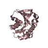
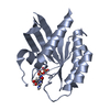
- Structure visualization
| Structure viewer | Molecule: MolmilJmol/JSmol |
|---|
- Downloads & links
-Download
| PDBx/mmCIF format | 6ukl.cif.gz | 114.3 KB | Display | PDBx/mmCIF format |
|---|---|---|---|---|
| PDB format | pdb6ukl.ent.gz | 86.1 KB | Display | PDB format |
| PDBx/mmJSON format | 6ukl.json.gz | Tree view | PDBx/mmJSON format | |
| Others |  Other downloads Other downloads |
-Validation report
| Arichive directory | https://data.pdbj.org/pub/pdb/validation_reports/uk/6uklftp://data.pdbj.org/pub/pdb/validation_reports/uk/6ukl | HTTPS FTP |
|---|
-Related structure data
| Related structure data |  6ukkC 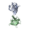 1qwdS S: Starting model for refinement C: citing same article ( |
|---|---|
| Similar structure data |


-Links
 PDBj
PDBj


- Assembly
| Deposited unit | 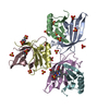
| ||||||||
|---|---|---|---|---|---|---|---|---|---|
| 1 | 
| ||||||||
| 2 | 
| ||||||||
| 3 | 
| ||||||||
| Unit cell |
| ||||||||
| Components on special symmetry positions |
|
-Components
-Outer membrane lipoprotein ... , 2 types, 6 molecules ACEDFB
| #1: Protein | Mass: 12298.773 Da / Num. of mol.: 3 / Fragment: N-terminal fragment (UNP residues 23-109) Source method: isolated from a genetically manipulated source Source: (gene. exp.) #2: Protein | Mass: 7902.951 Da / Num. of mol.: 3 / Fragment: C-terminal fragment (UNP residues 110-177) Source method: isolated from a genetically manipulated source Source: (gene. exp.) |
|---|
-Non-polymers , 4 types, 124 molecules 


| #3: Chemical | ChemComp-SO4 /  Mass: 96.063 Da / Num. of mol.: 15 / Source method: obtained synthetically / Formula: SO4 Mass: 96.063 Da / Num. of mol.: 15 / Source method: obtained synthetically / Formula: SO4#4: Chemical |  Mass: 195.237 Da / Num. of mol.: 2 / Source method: obtained synthetically / Formula: C6H13NO4S / Comment: pH buffer*YM Mass: 195.237 Da / Num. of mol.: 2 / Source method: obtained synthetically / Formula: C6H13NO4S / Comment: pH buffer*YM#5: Chemical | ChemComp-HEM / |  Mass: 616.487 Da / Num. of mol.: 1 / Source method: obtained synthetically / Formula: C34H32FeN4O4 Mass: 616.487 Da / Num. of mol.: 1 / Source method: obtained synthetically / Formula: C34H32FeN4O4#6: Water | ChemComp-HOH / | Mass: 18.015 Da / Num. of mol.: 106 / Source method: isolated from a natural source / Formula: H2O |
|---|
-Details
| Has ligand of interest | N |
|---|
-Experimental details
-Experiment
| Experiment | Method: X-RAY DIFFRACTION / Number of used crystals: 1 |
|---|
- Sample preparation
| Crystal | Density Matthews: 2.4 Å3/Da / Density % sol: 48.73 % |
|---|---|
| Crystal grow | Temperature: 294 K / Method: vapor diffusion, hanging drop / pH: 4.5 Details: 1.6 M ammonium sulfate, 0.1 M MES, pH 4.5, supplemented with 10% 0.1 M iron(III) chloride hexahydrate |
-Data collection
| Diffraction | Mean temperature: 100 K / Serial crystal experiment: N |
|---|---|
| Diffraction source | Source: SYNCHROTRON / Site: APS / Beamline: 21-ID-G / Wavelength: 0.97857 Å |
| Detector | Type: MARMOSAIC 300 mm CCD / Detector: CCD / Date: Nov 4, 2018 |
| Radiation | Monochromator: diamond(111) / Protocol: SINGLE WAVELENGTH / Monochromatic (M) / Laue (L): M / Scattering type: x-ray |
| Radiation wavelength | Wavelength: 0.97857 Å / Relative weight: 1 |
| Reflection | Resolution: 2.02→72.1 Å / Num. obs: 39425 / % possible obs: 100 % / Redundancy: 8.8 % / Biso Wilson estimate: 30.423 Å2 / Rpim(I) all: 0.033 / Rrim(I) all: 0.099 / Net I/σ(I): 11.5 / Num. measured all: 346459 |
| Reflection shell | Resolution: 2.02→2.05 Å / Redundancy: 7.9 % / Mean I/σ(I) obs: 2.1 / Num. unique obs: 1913 / Rpim(I) all: 0.335 / Rrim(I) all: 0.989 / % possible all: 97.5 |
-Phasing
| Phasing | Method: molecular replacement |
|---|
- Processing
| Software |
| |||||||||||||||||||||||||||||||||||||||||||||||||||||||||||||||||||||||||||
|---|---|---|---|---|---|---|---|---|---|---|---|---|---|---|---|---|---|---|---|---|---|---|---|---|---|---|---|---|---|---|---|---|---|---|---|---|---|---|---|---|---|---|---|---|---|---|---|---|---|---|---|---|---|---|---|---|---|---|---|---|---|---|---|---|---|---|---|---|---|---|---|---|---|---|---|---|
| Refinement | Method to determine structure: MOLECULAR REPLACEMENT Starting model: PDB entry 1QWD Resolution: 2.02→72.1 Å / Cor.coef. Fo:Fc: 0.954 / Cor.coef. Fo:Fc free: 0.939 / Cross valid method: THROUGHOUT / σ(F): 0 / ESU R: 0.18 / ESU R Free: 0.166 / Stereochemistry target values: MAXIMUM LIKELIHOOD Details: HYDROGENS HAVE BEEN ADDED IN THE RIDING POSITIONS U VALUES : REFINED INDIVIDUALLY
| |||||||||||||||||||||||||||||||||||||||||||||||||||||||||||||||||||||||||||
| Solvent computation | Ion probe radii: 0.8 Å / Shrinkage radii: 0.8 Å / VDW probe radii: 1.2 Å / Solvent model: MASK | |||||||||||||||||||||||||||||||||||||||||||||||||||||||||||||||||||||||||||
| Displacement parameters | Biso max: 127.69 Å2 / Biso mean: 36.833 Å2 / Biso min: 19.99 Å2
| |||||||||||||||||||||||||||||||||||||||||||||||||||||||||||||||||||||||||||
| Refinement step | Cycle: final / Resolution: 2.02→72.1 Å
| |||||||||||||||||||||||||||||||||||||||||||||||||||||||||||||||||||||||||||
| Refine LS restraints |
| |||||||||||||||||||||||||||||||||||||||||||||||||||||||||||||||||||||||||||
| LS refinement shell | Resolution: 2.02→2.072 Å / Total num. of bins used: 20
|
