 Movie
Movie Controller


+ Open data

- Basic information
| Entry | Database: PDB / ID: 2mat | ||||||
|---|---|---|---|---|---|---|---|
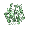
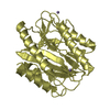
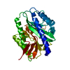
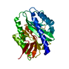
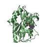
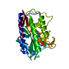
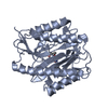
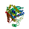
| Title | E.COLI METHIONINE AMINOPEPTIDASE AT 1.9 ANGSTROM RESOLUTION | ||||||
 Components Components | PROTEIN (METHIONINE AMINOPEPTIDASE) | ||||||
 Keywords Keywords | HYDROLASE / HYDROLASE(ALPHA-AMINOACYLPEPTIDE) | ||||||
| Function / homology |  Function and homology information Function and homology information: / methionyl aminopeptidase / initiator methionyl aminopeptidase activity / metalloaminopeptidase activity / ferrous iron binding / proteolysis / metal ion binding / cytosol Similarity search - Function | ||||||
| Biological species |  | ||||||
| Method |  X-RAY DIFFRACTION / MOLECULAR REPLACEMENT / Resolution: 1.9 Å X-RAY DIFFRACTION / MOLECULAR REPLACEMENT / Resolution: 1.9 Å | ||||||
 Authors Authors | Lowther, W.T. / Orville, A.M. / Madden, D.T. / Lim, S. / Rich, D.H. / Matthews, B.W. | ||||||
 Citation Citation | Journal: Biochemistry / Year: 1999 Title: Escherichia coli methionine aminopeptidase: implications of crystallographic analyses of the native, mutant, and inhibited enzymes for the mechanism of catalysis. Authors: Lowther, W.T. / Orville, A.M. / Madden, D.T. / Lim, S. / Rich, D.H. / Matthews, B.W. #1: Journal: Proc.Natl.Acad.Sci.USA / Year: 1998 Title: The anti-angiogenic agent fumagillin covalently modifies a conserved active-site histidine in the Escherichia coli methionine aminopeptidase. Authors: Lowther, W.T. / McMillen, D.A. / Orville, A.M. / Matthews, B.W. #2: Journal: Biochemistry / Year: 1993Title: Structure of the cobalt-dependent methionine aminopeptidase from Escherichia coli: a new type of proteolytic enzyme. Authors: Roderick, S.L. / Matthews, B.W. | ||||||
| History |
|
- Structure visualization
| Structure viewer | Molecule: MolmilJmol/JSmol |
|---|
- Downloads & links
-Download
| PDBx/mmCIF format | 2mat.cif.gz | 67.5 KB | Display | PDBx/mmCIF format |
|---|---|---|---|---|
| PDB format | pdb2mat.ent.gz | 47.8 KB | Display | PDB format |
| PDBx/mmJSON format | 2mat.json.gz | Tree view | PDBx/mmJSON format | |
| Others |  Other downloads Other downloads |
-Validation report
| Arichive directory | https://data.pdbj.org/pub/pdb/validation_reports/ma/2matftp://data.pdbj.org/pub/pdb/validation_reports/ma/2mat | HTTPS FTP |
|---|
-Related structure data
| Related structure data |  3matC  4matC  1matS S: Starting model for refinement C: citing same article ( |
|---|---|
| Similar structure data |
-Links
 PDBj
PDBj
- Assembly
| Deposited unit | 
| ||||||||
|---|---|---|---|---|---|---|---|---|---|
| 1 |
| ||||||||
| Unit cell |
|
-Components
| #1: Protein | Mass: 29341.775 Da / Num. of mol.: 1 / Mutation: R175Q Source method: isolated from a genetically manipulated source Details: SITE-DIRECTED MUTANT / Source: (gene. exp.) References: UniProt: P07906, UniProt: P0AE18*PLUS, methionyl aminopeptidase | ||||||
|---|---|---|---|---|---|---|---|
| #2: Chemical |   Mass: 58.933 Da / Num. of mol.: 3 / Source method: obtained synthetically / Formula: Co Mass: 58.933 Da / Num. of mol.: 3 / Source method: obtained synthetically / Formula: Co#3: Chemical | ChemComp-NA / |   Mass: 22.990 Da / Num. of mol.: 1 / Source method: obtained synthetically / Formula: Na Mass: 22.990 Da / Num. of mol.: 1 / Source method: obtained synthetically / Formula: Na#4: Water | ChemComp-HOH / |  Mass: 18.015 Da / Num. of mol.: 96 / Source method: isolated from a natural source / Formula: H2O Mass: 18.015 Da / Num. of mol.: 96 / Source method: isolated from a natural source / Formula: H2OSequence details | GLN 175, SITE-DIRECTED MUTANT | |
-Experimental details
-Experiment
| Experiment | Method: X-RAY DIFFRACTION / Number of used crystals: 1 |
|---|
- Sample preparation
| Crystal | Density Matthews: 2.05 Å3/Da / Density % sol: 39.4 % | ||||||||||||||||||||||||||||||||||||
|---|---|---|---|---|---|---|---|---|---|---|---|---|---|---|---|---|---|---|---|---|---|---|---|---|---|---|---|---|---|---|---|---|---|---|---|---|---|
| Crystal grow | Method: vapor diffusion, hanging drop / pH: 7.1 Details: CRYSTALS OF THE CO(II)-SUBSTITUTED ENZYME WERE GROWN AT ROOM TEMPERATURE BY VAPOR DIFFUSION IN 20-30 UL SITTING DROPS AFTER MIXING THE PROTEIN, 12 MG/ML SOLUTION IN STORAGE BUFFER(25 MM ...Details: CRYSTALS OF THE CO(II)-SUBSTITUTED ENZYME WERE GROWN AT ROOM TEMPERATURE BY VAPOR DIFFUSION IN 20-30 UL SITTING DROPS AFTER MIXING THE PROTEIN, 12 MG/ML SOLUTION IN STORAGE BUFFER(25 MM HEPES PH 6.8, 25 MM K2SO4, 100 MM NACL, 1 MM COCL2, 15 MM METHIONINE),CONTAINING 48.8 MM N-OCTANOYL SUCROSE, 1:1 WITH WELL SOLUTIONS (24-26% PEG4000, 0.1M HEPES PH7.0-7.2,FRESH 2 MM COCL2)., pH 7.1, VAPOR DIFFUSION, HANGING DROP | ||||||||||||||||||||||||||||||||||||
| Crystal grow | *PLUS Method: vapor diffusion, sitting dropDetails: drop consists of 1:1 mixture of well and protein solutions PH range low: 7.2 / PH range high: 7 | ||||||||||||||||||||||||||||||||||||
| Components of the solutions | *PLUS
|
-Data collection
| Diffraction | Mean temperature: 298 K |
|---|---|
| Diffraction source | Source: ROTATING ANODE / Type: RIGAKU RU200 / Wavelength: 1.5418 |
| Detector | Type: RIGAKU RAXIS IV / Detector: IMAGE PLATE / Date: Apr 10, 1998 |
| Radiation | Monochromator: NI FILTER / Protocol: SINGLE WAVELENGTH / Monochromatic (M) / Laue (L): M / Scattering type: x-ray |
| Radiation wavelength | Wavelength: 1.5418 Å / Relative weight: 1 |
| Reflection | Resolution: 1.9→35.5 Å / Num. obs: 18444 / % possible obs: 100 % / Redundancy: 5.1 % / Biso Wilson estimate: 15.5 Å2 / Rsym value: 0.077 / Net I/σ(I): 22.7 |
| Reflection shell | Resolution: 1.9→1.97 Å / Mean I/σ(I) obs: 6.1 / Rsym value: 0.251 / % possible all: 100 |
| Reflection | *PLUS Num. measured all: 94123 / Rmerge(I) obs: 0.077 |
| Reflection shell | *PLUS % possible obs: 100 % / Rmerge(I) obs: 0.251 |
- Processing
| Software |
| ||||||||||||||||||||||||||||||||||||||||||||||||||
|---|---|---|---|---|---|---|---|---|---|---|---|---|---|---|---|---|---|---|---|---|---|---|---|---|---|---|---|---|---|---|---|---|---|---|---|---|---|---|---|---|---|---|---|---|---|---|---|---|---|---|---|
| Refinement | Method to determine structure: MOLECULAR REPLACEMENT Starting model: 1MAT Resolution: 1.9→35.5 Å / Isotropic thermal model: TNT BCORREL V1.0 / σ(F): 0 / Stereochemistry target values: TNT PROTGEO
| ||||||||||||||||||||||||||||||||||||||||||||||||||
| Solvent computation | Solvent model: TNT / Bsol: 212.2 Å2 / ksol: 0.825 e/Å3 | ||||||||||||||||||||||||||||||||||||||||||||||||||
| Refinement step | Cycle: LAST / Resolution: 1.9→35.5 Å
| ||||||||||||||||||||||||||||||||||||||||||||||||||
| Refine LS restraints |
| ||||||||||||||||||||||||||||||||||||||||||||||||||
| Software | *PLUS Name: TNT / Version: 5F / Classification: refinement | ||||||||||||||||||||||||||||||||||||||||||||||||||
| Refine LS restraints | *PLUS Type: t_plane_restr / Dev ideal: 0.013 / Weight: 5 |
