 Movie
Movie Controller


+ Open data

- Basic information
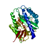
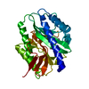
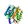
| Entry | Database: PDB / ID: 1c21 | ||||||
|---|---|---|---|---|---|---|---|
| Title | E. COLI METHIONINE AMINOPEPTIDASE: METHIONINE COMPLEX | ||||||
 Components Components | METHIONINE AMINOPEPTIDASE | ||||||
 Keywords Keywords | HYDROLASE / PRODUCT COMPLEX | ||||||
| Function / homology |  Function and homology information Function and homology information: / methionyl aminopeptidase / initiator methionyl aminopeptidase activity / metalloaminopeptidase activity / ferrous iron binding / proteolysis / metal ion binding / cytosol Similarity search - Function | ||||||
| Biological species |  | ||||||
| Method |  X-RAY DIFFRACTION / Resolution: 1.8 Å X-RAY DIFFRACTION / Resolution: 1.8 Å | ||||||
 Authors Authors | Lowther, W.T. / Zhang, Y. / Sampson, P.B. / Honek, J.F. / Matthews, B.W. | ||||||
 Citation Citation | Journal: Biochemistry / Year: 1999 Title: Insights into the mechanism of Escherichia coli methionine aminopeptidase from the structural analysis of reaction products and phosphorus-based transition-state analogues. Authors: Lowther, W.T. / Zhang, Y. / Sampson, P.B. / Honek, J.F. / Matthews, B.W. | ||||||
| History |
|
- Structure visualization
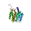
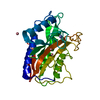
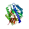
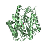
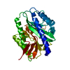
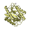
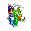
| Structure viewer | Molecule: MolmilJmol/JSmol |
|---|
- Downloads & links
-Download
| PDBx/mmCIF format | 1c21.cif.gz | 66.2 KB | Display | PDBx/mmCIF format |
|---|---|---|---|---|
| PDB format | pdb1c21.ent.gz | 47.4 KB | Display | PDB format |
| PDBx/mmJSON format | 1c21.json.gz | Tree view | PDBx/mmJSON format | |
| Others |  Other downloads Other downloads |
-Validation report
| Arichive directory | https://data.pdbj.org/pub/pdb/validation_reports/c2/1c21ftp://data.pdbj.org/pub/pdb/validation_reports/c2/1c21 | HTTPS FTP |
|---|
-Related structure data


-Links
 PDBj
PDBj
- Assembly
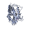
| Deposited unit | 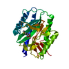
| ||||||||
|---|---|---|---|---|---|---|---|---|---|
| 1 |
| ||||||||
| Unit cell |
|
-Components
| #1: Protein | Mass: 29210.580 Da / Num. of mol.: 1 / Fragment: METHIONINE / Mutation: R175Q Source method: isolated from a genetically manipulated source Details: METHIONINE COMPLEX / Source: (gene. exp.) References: UniProt: P07906, UniProt: P0AE18*PLUS, methionyl aminopeptidase | ||||||||
|---|---|---|---|---|---|---|---|---|---|
| #2: Chemical |   Mass: 58.933 Da / Num. of mol.: 2 / Source method: obtained synthetically / Formula: Co Mass: 58.933 Da / Num. of mol.: 2 / Source method: obtained synthetically / Formula: Co#3: Chemical | ChemComp-NA / |   Mass: 22.990 Da / Num. of mol.: 1 / Source method: obtained synthetically / Formula: Na Mass: 22.990 Da / Num. of mol.: 1 / Source method: obtained synthetically / Formula: Na#4: Chemical | ChemComp-MET / |   Type: L-peptide linking / Mass: 149.211 Da / Num. of mol.: 1 / Source method: obtained synthetically / Formula: C5H11NO2S Type: L-peptide linking / Mass: 149.211 Da / Num. of mol.: 1 / Source method: obtained synthetically / Formula: C5H11NO2S#5: Water | ChemComp-HOH / |  Mass: 18.015 Da / Num. of mol.: 97 / Source method: isolated from a natural source / Formula: H2O Mass: 18.015 Da / Num. of mol.: 97 / Source method: isolated from a natural source / Formula: H2OSequence details | THERE ARE FOUR ADDITIONAL RESIDUES ON THE C-TERMINUS LEFT OVER FROM A THROMBIN DIGEST OF THE HIS- ...THERE ARE FOUR ADDITIONAL | |
-Experimental details
-Experiment
| Experiment | Method: X-RAY DIFFRACTION / Number of used crystals: 1 |
|---|
- Sample preparation
| Crystal | Density Matthews: 2.07 Å3/Da / Density % sol: 40.58 % | ||||||||||||||||||||||||||||||||||||||||||||||||||||||||||||||||||
|---|---|---|---|---|---|---|---|---|---|---|---|---|---|---|---|---|---|---|---|---|---|---|---|---|---|---|---|---|---|---|---|---|---|---|---|---|---|---|---|---|---|---|---|---|---|---|---|---|---|---|---|---|---|---|---|---|---|---|---|---|---|---|---|---|---|---|---|
| Crystal grow | Temperature: 298 K / Method: vapor diffusion, sitting drop Details: K2SO4, NaCl, methionine, N-octanoyl sucrose, PEG 4000, HEPES, CoCl2, VAPOR DIFFUSION, SITTING DROP, temperature 298K | ||||||||||||||||||||||||||||||||||||||||||||||||||||||||||||||||||
| Crystal grow | *PLUS pH: 6.8 | ||||||||||||||||||||||||||||||||||||||||||||||||||||||||||||||||||
| Components of the solutions | *PLUS
|
-Data collection
| Diffraction | Mean temperature: 298 K |
|---|---|
| Diffraction source | Source: ROTATING ANODE / Type: RIGAKU / Wavelength: 1.54 |
| Detector | Type: RIGAKU RAXIS / Detector: IMAGE PLATE / Date: Apr 23, 1999 |
| Radiation | Protocol: SINGLE WAVELENGTH / Monochromatic (M) / Laue (L): M / Scattering type: x-ray |
| Radiation wavelength | Wavelength: 1.54 Å / Relative weight: 1 |
| Reflection | Resolution: 1.8→24.8 Å / Num. all: 21097 / Num. obs: 85232 / % possible obs: 95.1 % / Redundancy: 4 % / Biso Wilson estimate: 19.4 Å2 / Rmerge(I) obs: 0.045 / Net I/σ(I): 35.2 |
| Reflection shell | Resolution: 1.8→1.86 Å / Rmerge(I) obs: 0.256 / Mean I/σ(I) obs: 4.4 / % possible all: 90.2 |
| Reflection | *PLUS Num. obs: 21097 / Num. measured all: 85232 |
| Reflection shell | *PLUS % possible obs: 90.2 % |
- Processing
| Software |
| |||||||||||||||
|---|---|---|---|---|---|---|---|---|---|---|---|---|---|---|---|---|
| Refinement | Resolution: 1.8→24.8 Å / Stereochemistry target values: TNT /
| |||||||||||||||
| Solvent computation | Solvent model: TNT / Bsol: 204.6 Å2 / ksol: 0.834 e/Å3 | |||||||||||||||
| Refinement step | Cycle: LAST / Resolution: 1.8→24.8 Å
| |||||||||||||||
| Refine LS restraints |
| |||||||||||||||
| Software | *PLUS Name: TNT / Classification: refinement | |||||||||||||||
| Refinement | *PLUS Highest resolution: 1.8 Å / Rfactor all: 0.163 / Num. reflection obs: 21097 | |||||||||||||||
| Solvent computation | *PLUS | |||||||||||||||
| Displacement parameters | *PLUS | |||||||||||||||
| Refine LS restraints | *PLUS
|
