[English] 日本語
 Yorodumi
- PDB-1mat: STRUCTURE OF THE COBALT-DEPENDENT METHIONINE AMINOPEPTIDASE FROM ... -
+ Open data
- Basic information
| Entry | Database: PDB / ID: 1mat | ||||||
|---|---|---|---|---|---|---|---|
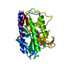
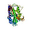
| Title | STRUCTURE OF THE COBALT-DEPENDENT METHIONINE AMINOPEPTIDASE FROM ESCHERICHIA COLI: A NEW TYPE OF PROTEOLYTIC ENZYME | ||||||
 Components Components | METHIONYL AMINOPEPTIDASE | ||||||
 Keywords Keywords | HYDROLASE(ALPHA-AMINOACYLPEPTIDE) | ||||||
| Function / homology |  Function and homology information Function and homology information: / methionyl aminopeptidase / initiator methionyl aminopeptidase activity / metalloaminopeptidase activity / ferrous iron binding / proteolysis / metal ion binding / cytosol Similarity search - Function | ||||||
| Biological species |  | ||||||
| Method |  X-RAY DIFFRACTION / Resolution: 2.4 Å X-RAY DIFFRACTION / Resolution: 2.4 Å | ||||||
 Authors Authors | Roderick, S.L. / Matthews, B.W. | ||||||
 Citation Citation |  Journal: Biochemistry / Year: 1993 Journal: Biochemistry / Year: 1993Title: Structure of the cobalt-dependent methionine aminopeptidase from Escherichia coli: a new type of proteolytic enzyme. Authors: Roderick, S.L. / Matthews, B.W. #1:  Journal: J.Biol.Chem. / Year: 1988 Journal: J.Biol.Chem. / Year: 1988Title: Crystallization of Methionine Aminopeptidase from Escherichia Coli Authors: Roderick, S.L. / Matthews, B.W. #2:  Journal: J.Bacteriol. / Year: 1987 Journal: J.Bacteriol. / Year: 1987Title: Processing of the Initiation Methionine from Proteins: Properties of the Escherichia Coli Methionine Aminopeptidase and its Gene Structure Authors: Ben-Bassat, A. / Bauer, K. / Chang, S.-Y. / Myambo, K. / Boosman, A. / Chang, S. | ||||||
| History |
| ||||||
| Remark 700 | SHEET THE SHEET STRUCTURE OF THIS MOLECULE IS BIFURCATED. IN ORDER TO REPRESENT THIS FEATURE IN THE ...SHEET THE SHEET STRUCTURE OF THIS MOLECULE IS BIFURCATED. IN ORDER TO REPRESENT THIS FEATURE IN THE SHEET RECORDS BELOW TWO SHEETS ARE DEFINED. STRANDS 2, 3, AND 4 OF 1A AND 1B ARE IDENTICAL. |
- Structure visualization
| Structure viewer | Molecule:  Molmil Molmil Jmol/JSmol Jmol/JSmol |
|---|
- Downloads & links
- Download
| PDBx/mmCIF format |  1mat.cif.gz 1mat.cif.gz | 62.2 KB | Display |  PDBx/mmCIF format PDBx/mmCIF format |
|---|---|---|---|---|
| PDB format |  pdb1mat.ent.gz pdb1mat.ent.gz | 45.3 KB | Display |  PDB format PDB format |
| PDBx/mmJSON format |  1mat.json.gz 1mat.json.gz | Tree view |  PDBx/mmJSON format PDBx/mmJSON format | |
| Others |  Other downloads Other downloads |
-Validation report
| Arichive directory |  https://data.pdbj.org/pub/pdb/validation_reports/ma/1mat https://data.pdbj.org/pub/pdb/validation_reports/ma/1mat ftp://data.pdbj.org/pub/pdb/validation_reports/ma/1mat ftp://data.pdbj.org/pub/pdb/validation_reports/ma/1mat | HTTPS FTP |
|---|
-Related structure data
| Similar structure data |
|---|
- Links
Links
- Assembly
| Deposited unit | 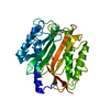
| ||||||||
|---|---|---|---|---|---|---|---|---|---|
| 1 |
| ||||||||
| Unit cell |
| ||||||||
| Atom site foot note | 1: CIS PROLINE - PRO 181 2: FOLLOWING RESIDUES TRUNCATED TO ALA: GLU 12, LYS 13, LYS 117, GLU 123, VAL 194, LEU 195, AND LYS 218. 3: RESIDUE LEU 88 TRUNCATED TO GLY. 4: FOLLOWING RESIDUES TRUNCATED TO GAMMA CARBON: GLU 9, ARG 93, LYS 86, LEU 87, LYS 89, THR 119, GLU 167, LYS 211, LYS 226, ARG 228, AND GLU 264. 5: FOLLOWING RESIDUES TRUNCATED TO DELTA CARBON: ARG 19, LYS 155, ARG 189, AND LYS 196. 6: RESIDUE LYS 252 TRUNCATED TO EPSILON CARBON. 7: FOLLOWING RESIDUES TRUNCATED TO VAL: ILE 49, ILE 120, AND ILE 144. | ||||||||
| Noncrystallographic symmetry (NCS) | NCS oper: (Code: given Matrix: (0.333795, 0.741892, 0.581532), Vector: Details | THE TRANSFORMATION PRESENTED ON *MTRIX* RECORDS BELOW WILL YIELD APPROXIMATE COORDINATES FOR PORTIONS OF THE CHAIN WHEN APPLIED TO OTHER PORTIONS OF THE CHAIN AS FOLLOWS: APPLYING MTRIX TO YIELDS ----------------- ----------------- THR 119 - MET 139 ILE 11 - TYR 31 VAL 140 - ILE 144 VAL 32 - VAL 36 ASN 145 - ALA 159 SER 37 - ASN 51 GLY 198 - ASN 208 GLY 91 - ILE 101 LEU 230 - THR 241 PHE 105 - GLY 116 | |
- Components
| #1: Protein | Mass: 29370.838 Da / Num. of mol.: 1 Source method: isolated from a genetically manipulated source Source: (gene. exp.)  References: UniProt: P07906, UniProt: P0AE18*PLUS, methionyl aminopeptidase | ||
|---|---|---|---|
| #2: Chemical | | #3: Water | ChemComp-HOH / | |
-Experimental details
-Experiment
| Experiment | Method:  X-RAY DIFFRACTION X-RAY DIFFRACTION |
|---|
- Sample preparation
| Crystal | Density Matthews: 2.13 Å3/Da / Density % sol: 42.27 % | ||||||||||||||||||||||||||||||||||||||||||||||||||||||
|---|---|---|---|---|---|---|---|---|---|---|---|---|---|---|---|---|---|---|---|---|---|---|---|---|---|---|---|---|---|---|---|---|---|---|---|---|---|---|---|---|---|---|---|---|---|---|---|---|---|---|---|---|---|---|---|
| Crystal grow | *PLUS pH: 6.8 / Method: vapor diffusion, hanging drop | ||||||||||||||||||||||||||||||||||||||||||||||||||||||
| Components of the solutions | *PLUS
|
-Data collection
| Radiation | Scattering type: x-ray |
|---|---|
| Radiation wavelength | Relative weight: 1 |
| Reflection | *PLUS Highest resolution: 2.5 Å / Num. obs: 8054 / % possible obs: 93 % / Rmerge(I) obs: 0.035 |
- Processing
| Software | Name: TNT / Classification: refinement | ||||||||||||||||||||||||||||||
|---|---|---|---|---|---|---|---|---|---|---|---|---|---|---|---|---|---|---|---|---|---|---|---|---|---|---|---|---|---|---|---|
| Refinement | Rfactor obs: 0.182 / Highest resolution: 2.4 Å | ||||||||||||||||||||||||||||||
| Refinement step | Cycle: LAST / Highest resolution: 2.4 Å
| ||||||||||||||||||||||||||||||
| Refine LS restraints |
| ||||||||||||||||||||||||||||||
| Software | *PLUS Name: TNT / Classification: refinement | ||||||||||||||||||||||||||||||
| Refinement | *PLUS Highest resolution: 2.5 Å / Lowest resolution: 52 Å / Num. reflection obs: 8387 / Rfactor obs: 0.182 | ||||||||||||||||||||||||||||||
| Solvent computation | *PLUS | ||||||||||||||||||||||||||||||
| Displacement parameters | *PLUS | ||||||||||||||||||||||||||||||
| Refine LS restraints | *PLUS
|
 Movie
Movie Controller





 PDBj
PDBj


