 Movie
Movie Controller

[English] 日本語
 Yorodumi
- PDB-3tb5: Crystal Structure of the Enterococcus faecalis Methionine aminope... -
+ Open data

- Basic information
| Entry | Database: PDB / ID: 3tb5 | ||||||
|---|---|---|---|---|---|---|---|
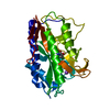
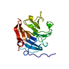
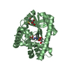
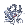
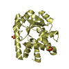
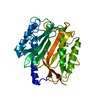
| Title | Crystal Structure of the Enterococcus faecalis Methionine aminopeptidase apo form | ||||||
 Components Components | Methionine aminopeptidase | ||||||
 Keywords Keywords | HYDROLASE / Methionine aminopeptidase / metalloprotease / enterococcus feacalis | ||||||
| Function / homology | Creatine Amidinohydrolase / Creatinase/methionine aminopeptidase superfamily / Alpha-Beta Complex / Alpha Beta / CITRIC ACID / :  Function and homology information Function and homology information | ||||||
| Biological species |   Enterococcus faecalis (bacteria) Enterococcus faecalis (bacteria) | ||||||
| Method |  X-RAY DIFFRACTION / SYNCHROTRON / MOLECULAR REPLACEMENT / Resolution: 2.3 Å X-RAY DIFFRACTION / SYNCHROTRON / MOLECULAR REPLACEMENT / Resolution: 2.3 Å | ||||||
 Authors Authors | Kishor, C. / Gumpena, R. / Reddi, R. / Addlagatta, A. | ||||||
 Citation Citation | Journal: MEDCHEMCOMM / Year: 2012 Title: Structural studies of Enterococcus faecalis methionine aminopeptidase and design of microbe specific 2,2'-bipyridine based inhibitors Authors: Kishor, C. / Gumpena, R. / Reddi, R. / Addlagatta, A. | ||||||
| History |
|
- Structure visualization
| Structure viewer | Molecule: MolmilJmol/JSmol |
|---|
- Downloads & links
-Download
| PDBx/mmCIF format | 3tb5.cif.gz | 154.5 KB | Display | PDBx/mmCIF format |
|---|---|---|---|---|
| PDB format | pdb3tb5.ent.gz | 123.4 KB | Display | PDB format |
| PDBx/mmJSON format | 3tb5.json.gz | Tree view | PDBx/mmJSON format | |
| Others |  Other downloads Other downloads |
-Validation report
| Arichive directory | https://data.pdbj.org/pub/pdb/validation_reports/tb/3tb5ftp://data.pdbj.org/pub/pdb/validation_reports/tb/3tb5 | HTTPS FTP |
|---|
-Related structure data
| Related structure data |  1c21S S: Starting model for refinement |
|---|---|
| Similar structure data |


-Links
 PDBj
PDBj- Assembly
| Deposited unit | 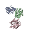
| ||||||||
|---|---|---|---|---|---|---|---|---|---|
| 1 | 
| ||||||||
| 2 | 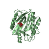
| ||||||||
| 3 | 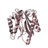
| ||||||||
| Unit cell |
|
-Components
| #1: Protein | Mass: 29449.436 Da / Num. of mol.: 3 Source method: isolated from a genetically manipulated source Source: (gene. exp.) Enterococcus faecalis (bacteria) / Strain: HIP11704 / Gene: EFHG_00941, MetAP / Plasmid: pET28a / Production host: #2: Chemical |   Mass: 192.124 Da / Num. of mol.: 2 / Source method: obtained synthetically / Formula: C6H8O7 Mass: 192.124 Da / Num. of mol.: 2 / Source method: obtained synthetically / Formula: C6H8O7#3: Water | ChemComp-HOH / |  Mass: 18.015 Da / Num. of mol.: 159 / Source method: isolated from a natural source / Formula: H2O Mass: 18.015 Da / Num. of mol.: 159 / Source method: isolated from a natural source / Formula: H2O |
|---|
-Experimental details
-Experiment
| Experiment | Method: X-RAY DIFFRACTION / Number of used crystals: 4 |
|---|
- Sample preparation
| Crystal | Density Matthews: 2.8 Å3/Da / Density % sol: 56.07 % |
|---|---|
| Crystal grow | Temperature: 298 K / Method: vapor diffusion, hanging drop / pH: 5.6 Details: 14-20% PEG 3400, 0.14M (NH4)2H-citrate, 8% glycerol, pH 5.4-6.2, VAPOR DIFFUSION, HANGING DROP, temperature 298K |
-Data collection
| Diffraction | Mean temperature: 100 K |
|---|---|
| Diffraction source | Source: SYNCHROTRON / Site: ALS  / Beamline: 8.2.2 / Wavelength: 0.9919 Å / Beamline: 8.2.2 / Wavelength: 0.9919 Å |
| Detector | Type: ADSC QUANTUM 315 / Detector: CCD / Date: Mar 25, 2011 |
| Radiation | Protocol: SINGLE WAVELENGTH / Monochromatic (M) / Laue (L): M / Scattering type: x-ray |
| Radiation wavelength | Wavelength: 0.9919 Å / Relative weight: 1 |
| Reflection | Resolution: 2.3→45.22 Å / Num. obs: 43107 / % possible obs: 99 % / Observed criterion σ(F): 0 / Observed criterion σ(I): 0 / Redundancy: 3.5 % / Rmerge(I) obs: 0.052 / Rsym value: 0.033 |
- Processing
| Software |
| ||||||||||||||||||||||||||||||||||||||||||||||||||||||||||||||||||||||||||||||||||||||||||||||||||||||||||||||||||||||||||||||||||||||||||||||||||||||||||||||||||||||||||
|---|---|---|---|---|---|---|---|---|---|---|---|---|---|---|---|---|---|---|---|---|---|---|---|---|---|---|---|---|---|---|---|---|---|---|---|---|---|---|---|---|---|---|---|---|---|---|---|---|---|---|---|---|---|---|---|---|---|---|---|---|---|---|---|---|---|---|---|---|---|---|---|---|---|---|---|---|---|---|---|---|---|---|---|---|---|---|---|---|---|---|---|---|---|---|---|---|---|---|---|---|---|---|---|---|---|---|---|---|---|---|---|---|---|---|---|---|---|---|---|---|---|---|---|---|---|---|---|---|---|---|---|---|---|---|---|---|---|---|---|---|---|---|---|---|---|---|---|---|---|---|---|---|---|---|---|---|---|---|---|---|---|---|---|---|---|---|---|---|---|---|---|
| Refinement | Method to determine structure: MOLECULAR REPLACEMENT Starting model: PDB ENTRY 1C21 Resolution: 2.3→36.47 Å / Cor.coef. Fo:Fc: 0.939 / Cor.coef. Fo:Fc free: 0.895 / SU B: 5.834 / SU ML: 0.146 / Cross valid method: THROUGHOUT / ESU R: 0.269 / ESU R Free: 0.226 / Stereochemistry target values: MAXIMUM LIKELIHOOD / Details: HYDROGENS HAVE BEEN ADDED IN THE RIDING POSITIONS
| ||||||||||||||||||||||||||||||||||||||||||||||||||||||||||||||||||||||||||||||||||||||||||||||||||||||||||||||||||||||||||||||||||||||||||||||||||||||||||||||||||||||||||
| Solvent computation | Ion probe radii: 0.8 Å / Shrinkage radii: 0.8 Å / VDW probe radii: 1.4 Å / Solvent model: MASK | ||||||||||||||||||||||||||||||||||||||||||||||||||||||||||||||||||||||||||||||||||||||||||||||||||||||||||||||||||||||||||||||||||||||||||||||||||||||||||||||||||||||||||
| Displacement parameters | Biso mean: 37.177 Å2
| ||||||||||||||||||||||||||||||||||||||||||||||||||||||||||||||||||||||||||||||||||||||||||||||||||||||||||||||||||||||||||||||||||||||||||||||||||||||||||||||||||||||||||
| Refinement step | Cycle: LAST / Resolution: 2.3→36.47 Å
| ||||||||||||||||||||||||||||||||||||||||||||||||||||||||||||||||||||||||||||||||||||||||||||||||||||||||||||||||||||||||||||||||||||||||||||||||||||||||||||||||||||||||||
| Refine LS restraints |
| ||||||||||||||||||||||||||||||||||||||||||||||||||||||||||||||||||||||||||||||||||||||||||||||||||||||||||||||||||||||||||||||||||||||||||||||||||||||||||||||||||||||||||
| LS refinement shell | Resolution: 2.3→2.36 Å / Total num. of bins used: 20
|
