Mass: 17900.031 Da / Num. of mol.: 1 / Fragment: calcium binding domain 2 Source method: isolated from a genetically manipulated source Source: (gene. exp.) Mus musculus (house mouse) / Gene: Slc8a3 / Plasmid: pET23b / Production host: Escherichia coli (E. coli) / Strain (production host): BL21(DE3) / References: UniProt: Q7TS90, UniProt: S4R2P9*PLUS
-
Experimental details
-
Experiment
Experiment
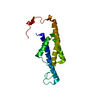
Method: SOLUTION NMR Details: The solution structure of calcium binding domain 2B of NCX3 is solved in excess of 10 mM CaCl2. However, Ca ions are not modeled in the structure, because resonances of crucial coordinating ...Details: The solution structure of calcium binding domain 2B of NCX3 is solved in excess of 10 mM CaCl2. However, Ca ions are not modeled in the structure, because resonances of crucial coordinating residues are lacking: res 605 to 610 and 665 to 669.
NMR experiment
Conditions-ID
Experiment-ID
Solution-ID
Type
1
1
1
2D 1H-15N HSQC
1
2
1
3D HNCO
1
3
1
3D HN(CA)CB
1
4
1
3DCBCA(CO)NH
1
5
1
3DHBHA(CO)NH
1
6
1
3D (H)CCH-TOCSY
1
7
1
3D HNHA
1
8
2
2D 1H-15N IPAPHSQC
1
9
3
2D 1H-13C IPAPHSQC
1
10
1
3D 1H-13C NOESY aliphatic
1
11
1
3D 1H-13C NOESY aromatic
1
12
1
3D 1H-15N NOESY
1
13
1
3DC(CO)NH
-
Sample preparation
Details
Solution-ID
Contents
Solvent system
1
0.5 mM [U-100% 13C; U-100% 15N] protein, 20 mM HEPES, 20 mM beta-mercaptoethanol, 10 mM CaCl2, 95% H2O/5% D2O
95% H2O/5% D2O
2
0.5 mM [U-100% 15N] protein, 20 mM HEPES, 20 mM beta-mercaptoethanol, 10 mM CaCl2, 8 mg/mL Pf1 phage, 95% H2O/5% D2O
95% H2O/5% D2O
3
0.5 mM [U-100% 15N] protein, 20 mM HEPES, 20 mM beta-mercaptoethanol, 10 mM CaCl2, 95% H2O/5% D2O
Method: simulated annealing / Software ordinal: 1 Details: THE SOLUTION STRUCTURE OF CALCIUM BINDING DOMAIN 2B OF NCX3 IS SOLVED IN EXCESS OF 10 MM CACL2. HOWEVER, CA IONS ARE NOT MODELED IN THE STRUCTURE, BECAUSE RESONANCES OF CRUCIAL COORDINATING ...Details: THE SOLUTION STRUCTURE OF CALCIUM BINDING DOMAIN 2B OF NCX3 IS SOLVED IN EXCESS OF 10 MM CACL2. HOWEVER, CA IONS ARE NOT MODELED IN THE STRUCTURE, BECAUSE RESONANCES OF CRUCIAL COORDINATING RESIDUES ARE LACKING: RES 605 TO 610 AND 665 TO 669. THE REFINEMENT PROTOCOL IN EXPLICIT WATER, INCLUDING DISTANCE AND DIHEDRAL RESTRAINTS. THE RDC RESTRAINTS WERE NOT INCLUDED IN THE REFINEMENT.
NMR constraints
NOE constraints total: 2901 / NOE long range total count: 1289 / NOE medium range total count: 279 / Protein phi angle constraints total count: 93 / Protein psi angle constraints total count: 93
NMR representative
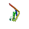
Selection criteria: lowest energy
NMR ensemble
Conformer selection criteria: target function / Conformers calculated total number: 100 / Conformers submitted total number: 20
+
About Yorodumi
-
News
-
Feb 9, 2022. New format data for meta-information of EMDB entries
New format data for meta-information of EMDB entries
Version 3 of the EMDB header file is now the official format.
The previous official version 1.9 will be removed from the archive.
In the structure databanks used in Yorodumi, some data are registered as the other names, "COVID-19 virus" and "2019-nCoV". Here are the details of the virus and the list of structure data.
Jan 31, 2019. EMDB accession codes are about to change! (news from PDBe EMDB page)
EMDB accession codes are about to change! (news from PDBe EMDB page)
The allocation of 4 digits for EMDB accession codes will soon come to an end. Whilst these codes will remain in use, new EMDB accession codes will include an additional digit and will expand incrementally as the available range of codes is exhausted. The current 4-digit format prefixed with “EMD-” (i.e. EMD-XXXX) will advance to a 5-digit format (i.e. EMD-XXXXX), and so on. It is currently estimated that the 4-digit codes will be depleted around Spring 2019, at which point the 5-digit format will come into force.
The EM Navigator/Yorodumi systems omit the EMD- prefix.
Related info.:Q: What is EMD? / ID/Accession-code notation in Yorodumi/EM Navigator
Yorodumi is a browser for structure data from EMDB, PDB, SASBDB, etc.
This page is also the successor to EM Navigator detail page, and also detail information page/front-end page for Omokage search.
The word "yorodu" (or yorozu) is an old Japanese word meaning "ten thousand". "mi" (miru) is to see.
Related info.:EMDB / PDB / SASBDB / Comparison of 3 databanks / Yorodumi Search / Aug 31, 2016. New EM Navigator & Yorodumi / Yorodumi Papers / Jmol/JSmol / Function and homology information / Changes in new EM Navigator and Yorodumi
 Movie
Movie Controller

 Yorodumi
 Open data
 Basic information
 Components
 Keywords
 Function and homology information
 Authors
 Citation
 Structure visualization
 Downloads & links
 Other downloads
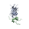


 PDBj
PDBj


 Assembly
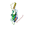

 HSQC
HSQC Sample preparation
 Processing