 Movie
Movie Controller

[English] 日本語
 Yorodumi
- PDB-1jbg: Crystal Structure of MtaN, the Bacillus subtilis Multidrug Transp... -
+ Open data

- Basic information
| Entry | Database: PDB / ID: 1jbg | ||||||
|---|---|---|---|---|---|---|---|
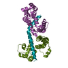
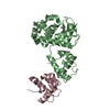
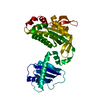
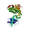
| Title | Crystal Structure of MtaN, the Bacillus subtilis Multidrug Transporter Activator, N-terminus | ||||||
 Components Components | transcription activator of multidrug-efflux transporter genes mta | ||||||
 Keywords Keywords | TRANSCRIPTION / winged helix-turn-helix / antiparallel coiled-coil | ||||||
| Function / homology |  Function and homology information Function and homology informationDNA-binding transcription factor activity / regulation of DNA-templated transcription / DNA binding / cytoplasm Similarity search - Function | ||||||
| Biological species |  | ||||||
| Method |  X-RAY DIFFRACTION / SYNCHROTRON / MAD / Resolution: 2.75 Å X-RAY DIFFRACTION / SYNCHROTRON / MAD / Resolution: 2.75 Å | ||||||
 Authors Authors | Godsey, M.H. / Neyfakh, A.A. / Brennan, R.G. | ||||||
 Citation Citation | Journal: J.Biol.Chem. / Year: 2001 Title: Crystal structure of MtaN, a global multidrug transporter gene activator. Authors: Godsey, M.H. / Baranova, N.N. / Neyfakh, A.A. / Brennan, R.G. | ||||||
| History |
|
- Structure visualization
| Structure viewer | Molecule: MolmilJmol/JSmol |
|---|
- Downloads & links
-Download
| PDBx/mmCIF format | 1jbg.cif.gz | 32.8 KB | Display | PDBx/mmCIF format |
|---|---|---|---|---|
| PDB format | pdb1jbg.ent.gz | 22.7 KB | Display | PDB format |
| PDBx/mmJSON format | 1jbg.json.gz | Tree view | PDBx/mmJSON format | |
| Others |  Other downloads Other downloads |
-Validation report
| Arichive directory | https://data.pdbj.org/pub/pdb/validation_reports/jb/1jbgftp://data.pdbj.org/pub/pdb/validation_reports/jb/1jbg | HTTPS FTP |
|---|
-Related structure data
| Similar structure data |
|---|


-Links
 PDBj
PDBj

- Assembly
| Deposited unit | 
| ||||||||
|---|---|---|---|---|---|---|---|---|---|
| 1 | 
| ||||||||
| Unit cell |
| ||||||||
| Details | The other half of the biological assembly is generated by: -X+1, 1/2-Y+1, Z. |
-Components
| #1: Protein | Mass: 12861.807 Da / Num. of mol.: 1 / Fragment: N-terminus (Residues 1-109) Source method: isolated from a genetically manipulated source Source: (gene. exp.) |
|---|---|
| #2: Water | ChemComp-HOH /  Mass: 18.015 Da / Num. of mol.: 16 / Source method: isolated from a natural source / Formula: H2O Mass: 18.015 Da / Num. of mol.: 16 / Source method: isolated from a natural source / Formula: H2O |
-Experimental details
-Experiment
| Experiment | Method: X-RAY DIFFRACTION / Number of used crystals: 1 |
|---|
- Sample preparation
| Crystal | Density Matthews: 3.74 Å3/Da / Density % sol: 67.14 % | |||||||||||||||||||||||||||||||||||
|---|---|---|---|---|---|---|---|---|---|---|---|---|---|---|---|---|---|---|---|---|---|---|---|---|---|---|---|---|---|---|---|---|---|---|---|---|
| Crystal grow | Temperature: 298 K / Method: vapor diffusion, hanging drop / pH: 7 Details: lithium chloride, magnesium chloride, HEPES, pH 7.0, VAPOR DIFFUSION, HANGING DROP, temperature 298.0K | |||||||||||||||||||||||||||||||||||
| Crystal grow | *PLUS Details: Godsey, M., (2000) Acta Crystallogr., D56, 1456. | |||||||||||||||||||||||||||||||||||
| Components of the solutions | *PLUS
|
-Data collection
| Diffraction | Mean temperature: 100 K |
|---|---|
| Diffraction source | Source: SYNCHROTRON / Site: SSRL  / Beamline: BL7-1 / Wavelength: 1.08 Å / Beamline: BL7-1 / Wavelength: 1.08 Å |
| Detector | Type: MARRESEARCH / Detector: IMAGE PLATE / Date: Dec 13, 1999 |
| Radiation | Monochromator: 58 cm long, Pt-coated, fused silica, vertical focus mirror; Cyclindrically bent triangular Si(111) asymmetric cut, horizontal focus monochromator Protocol: SINGLE WAVELENGTH / Monochromatic (M) / Laue (L): M / Scattering type: x-ray |
| Radiation wavelength | Wavelength: 1.08 Å / Relative weight: 1 |
| Reflection | Resolution: 2.75→23.3 Å / Num. all: 5246 / Num. obs: 5199 / % possible obs: 99.1 % / Observed criterion σ(F): 0 / Observed criterion σ(I): 0 / Redundancy: 8.1 % / Biso Wilson estimate: 121.3 Å2 / Rmerge(I) obs: 0.072 / Rsym value: 0.072 / Net I/σ(I): 7.9 |
| Reflection shell | Resolution: 2.75→2.82 Å / Redundancy: 4.8 % / Rmerge(I) obs: 0.271 / Mean I/σ(I) obs: 2.7 / Num. unique all: 377 / Rsym value: 0.271 / % possible all: 99 |
| Reflection | *PLUS % possible obs: 99.3 % / Num. measured all: 42297 |
| Reflection shell | *PLUS % possible obs: 99 % |
- Processing
| Software |
| ||||||||||||||||||||||||||||||||||||||||
|---|---|---|---|---|---|---|---|---|---|---|---|---|---|---|---|---|---|---|---|---|---|---|---|---|---|---|---|---|---|---|---|---|---|---|---|---|---|---|---|---|---|
| Refinement | Method to determine structure: MAD / Resolution: 2.75→23.33 Å / Rfactor Rfree error: 0.018 / Data cutoff high absF: 157528.71 / Data cutoff low absF: 0 / Isotropic thermal model: ANISOTROPIC, RESTRAINED / Cross valid method: THROUGHOUT / σ(F): 0 / σ(I): 0 / Stereochemistry target values: Engh & Huber
| ||||||||||||||||||||||||||||||||||||||||
| Solvent computation | Solvent model: FLAT MODEL / Bsol: 43.17 Å2 / ksol: 0.326 e/Å3 | ||||||||||||||||||||||||||||||||||||||||
| Displacement parameters | Biso mean: 81.6 Å2
| ||||||||||||||||||||||||||||||||||||||||
| Refine analyze |
| ||||||||||||||||||||||||||||||||||||||||
| Refinement step | Cycle: LAST / Resolution: 2.75→23.33 Å
| ||||||||||||||||||||||||||||||||||||||||
| Refine LS restraints |
| ||||||||||||||||||||||||||||||||||||||||
| LS refinement shell | Resolution: 2.75→2.92 Å / Rfactor Rfree error: 0.088 / Total num. of bins used: 6
| ||||||||||||||||||||||||||||||||||||||||
| Xplor file |
| ||||||||||||||||||||||||||||||||||||||||
| Software | *PLUS Name: CNS / Version: 1 / Classification: refinement | ||||||||||||||||||||||||||||||||||||||||
| Refinement | *PLUS Lowest resolution: 25 Å / σ(F): 0 / % reflection Rfree: 5.4 % / Rfactor obs: 0.228 | ||||||||||||||||||||||||||||||||||||||||
| Solvent computation | *PLUS | ||||||||||||||||||||||||||||||||||||||||
| Displacement parameters | *PLUS Biso mean: 81.6 Å2 | ||||||||||||||||||||||||||||||||||||||||
| Refine LS restraints | *PLUS
| ||||||||||||||||||||||||||||||||||||||||
| LS refinement shell | *PLUS Rfactor Rfree: 0.527 / % reflection Rfree: 5.2 % / Rfactor Rwork: 0.4 |
