 Movie
Movie Controller


+ Open data

- Basic information
| Entry | Database: PDB / ID: 4ug3 | ||||||
|---|---|---|---|---|---|---|---|
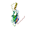
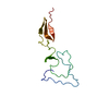
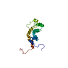
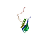
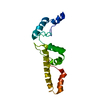
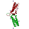
| Title | B. subtilis GpsB N-terminal Domain | ||||||
 Components Components | CELL CYCLE PROTEIN GPSB | ||||||
 Keywords Keywords | CELL CYCLE / BACTERIAL GROWTH REGULATION / CELL WALL SYNTHESIS / CELL DIVISION | ||||||
| Function / homology | Cell cycle protein GpsB / DivIVA family / DivIVA domain / DivIVA protein / peptidoglycan-based cell wall biogenesis / regulation of cell shape / cell division / cytoplasm / Cell cycle adapter protein GpsB Function and homology information Function and homology information | ||||||
| Biological species |  | ||||||
| Method |  X-RAY DIFFRACTION / SYNCHROTRON / MOLECULAR REPLACEMENT / Resolution: 2.8 Å X-RAY DIFFRACTION / SYNCHROTRON / MOLECULAR REPLACEMENT / Resolution: 2.8 Å | ||||||
 Authors Authors | Rismondo, J. / Cleverley, R.M. / Lane, H.V. / Grohennig, S. / Steglich, A. / Moller, L. / Krishna Mannala, G. / Hain, T. / Lewis, R.J. / Halbedel, S. | ||||||
 Citation Citation | Journal: Mol.Microbiol. / Year: 2016 Title: Structure of the Bacterial Cell Division Determinant Gpsb and its Interaction with Penicillin Binding Proteins. Authors: Rismondo, J. / Cleverley, R.M. / Lane, H.V. / Grosshennig, S. / Steglich, A. / Moller, L. / Mannala, G.K. / Hain, T. / Lewis, R.J. / Halbedel, S. | ||||||
| History |
|
- Structure visualization
| Structure viewer | Molecule: MolmilJmol/JSmol |
|---|
- Downloads & links
-Download
| PDBx/mmCIF format | 4ug3.cif.gz | 58.8 KB | Display | PDBx/mmCIF format |
|---|---|---|---|---|
| PDB format | pdb4ug3.ent.gz | 44.2 KB | Display | PDB format |
| PDBx/mmJSON format | 4ug3.json.gz | Tree view | PDBx/mmJSON format | |
| Others |  Other downloads Other downloads |
-Validation report
| Arichive directory | https://data.pdbj.org/pub/pdb/validation_reports/ug/4ug3ftp://data.pdbj.org/pub/pdb/validation_reports/ug/4ug3 | HTTPS FTP |
|---|
-Related structure data
| Related structure data | 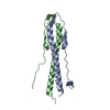 4ug1SC 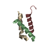 5an5C S: Starting model for refinement C: citing same article ( |
|---|---|
| Similar structure data |
-Links
 PDBj
PDBj- Assembly
| Deposited unit | 
| ||||||||
|---|---|---|---|---|---|---|---|---|---|
| 1 | 
| ||||||||
| 2 | 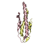
| ||||||||
| Unit cell |
|
-Components
| #1: Protein | Mass: 8413.601 Da / Num. of mol.: 4 / Fragment: N-TERMINAL DOMAIN, UNP RESIDUES 1-68 Source method: isolated from a genetically manipulated source Source: (gene. exp.) #2: Water | ChemComp-HOH / |  Mass: 18.015 Da / Num. of mol.: 2 / Source method: isolated from a natural source / Formula: H2O Mass: 18.015 Da / Num. of mol.: 2 / Source method: isolated from a natural source / Formula: H2O |
|---|
-Experimental details
-Experiment
| Experiment | Method: X-RAY DIFFRACTION / Number of used crystals: 1 |
|---|
- Sample preparation
| Crystal | Density Matthews: 2.31 Å3/Da / Density % sol: 46.72 % / Description: NONE |
|---|---|
| Crystal grow | pH: 6.5 Details: 0.05M BIS TRIS PH 6.5, 0.05M AMMONIUM SULPHATE, 30% PENTAERYTHRITOL ETHOXYLATE |
-Data collection
| Diffraction | Mean temperature: 100 K |
|---|---|
| Diffraction source | Source: SYNCHROTRON / Site: Diamond  / Beamline: I04 / Wavelength: 0.97949 / Beamline: I04 / Wavelength: 0.97949 |
| Detector | Type: DECTRIS PILATUS 6M / Detector: PIXEL / Date: Feb 14, 2015 |
| Radiation | Monochromator: SI (111) DOUBLE CRYSTAL MONOCHROMATOR / Protocol: SINGLE WAVELENGTH / Monochromatic (M) / Laue (L): M / Scattering type: x-ray |
| Radiation wavelength | Wavelength: 0.97949 Å / Relative weight: 1 |
| Reflection | Resolution: 2.8→50.3 Å / Num. obs: 7791 / % possible obs: 100 % / Observed criterion σ(I): 0 / Redundancy: 3.7 % / Rmerge(I) obs: 0.1 / Net I/σ(I): 10.6 |
| Reflection shell | Resolution: 2.8→2.95 Å / Redundancy: 3.6 % / Rmerge(I) obs: 0.49 / Mean I/σ(I) obs: 3.7 / % possible all: 99.9 |
- Processing
| Software |
| ||||||||||||||||||||||||||||||||||||||||||||||||||||||||||||||||||||||||||||||||||||||||||||||||||||||||||||||||||||||||||||||||||||||||||||||||||||||||||||||||||||||||||||||||||||||
|---|---|---|---|---|---|---|---|---|---|---|---|---|---|---|---|---|---|---|---|---|---|---|---|---|---|---|---|---|---|---|---|---|---|---|---|---|---|---|---|---|---|---|---|---|---|---|---|---|---|---|---|---|---|---|---|---|---|---|---|---|---|---|---|---|---|---|---|---|---|---|---|---|---|---|---|---|---|---|---|---|---|---|---|---|---|---|---|---|---|---|---|---|---|---|---|---|---|---|---|---|---|---|---|---|---|---|---|---|---|---|---|---|---|---|---|---|---|---|---|---|---|---|---|---|---|---|---|---|---|---|---|---|---|---|---|---|---|---|---|---|---|---|---|---|---|---|---|---|---|---|---|---|---|---|---|---|---|---|---|---|---|---|---|---|---|---|---|---|---|---|---|---|---|---|---|---|---|---|---|---|---|---|---|
| Refinement | Method to determine structure: MOLECULAR REPLACEMENT Starting model: PDB ENTRY 4UG1 Resolution: 2.8→50.3 Å / Cor.coef. Fo:Fc: 0.931 / Cor.coef. Fo:Fc free: 0.848 / SU B: 17.384 / SU ML: 0.338 / Cross valid method: THROUGHOUT / ESU R Free: 0.462 / Stereochemistry target values: MAXIMUM LIKELIHOOD Details: HYDROGENS HAVE BEEN ADDED IN THE RIDING POSITIONS. U VALUES REFINED INDIVIDUALLY
| ||||||||||||||||||||||||||||||||||||||||||||||||||||||||||||||||||||||||||||||||||||||||||||||||||||||||||||||||||||||||||||||||||||||||||||||||||||||||||||||||||||||||||||||||||||||
| Solvent computation | Ion probe radii: 0.8 Å / Shrinkage radii: 0.8 Å / VDW probe radii: 1.2 Å / Solvent model: MASK | ||||||||||||||||||||||||||||||||||||||||||||||||||||||||||||||||||||||||||||||||||||||||||||||||||||||||||||||||||||||||||||||||||||||||||||||||||||||||||||||||||||||||||||||||||||||
| Displacement parameters | Biso mean: 53.503 Å2
| ||||||||||||||||||||||||||||||||||||||||||||||||||||||||||||||||||||||||||||||||||||||||||||||||||||||||||||||||||||||||||||||||||||||||||||||||||||||||||||||||||||||||||||||||||||||
| Refinement step | Cycle: LAST / Resolution: 2.8→50.3 Å
| ||||||||||||||||||||||||||||||||||||||||||||||||||||||||||||||||||||||||||||||||||||||||||||||||||||||||||||||||||||||||||||||||||||||||||||||||||||||||||||||||||||||||||||||||||||||
| Refine LS restraints |
|