[English] 日本語
Yorodumi
- PDB-2l2p: Folding Intermediate of the Fyn SH3 A39V/N53P/V55L from NMR Relax... -

+
Open data


ID or keywords:

Loading...

-
Basic information

Entry
Database: PDB / ID: 2l2p
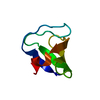
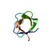
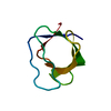
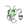
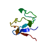
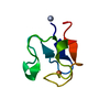
TitleFolding Intermediate of the Fyn SH3 A39V/N53P/V55L from NMR Relaxation Dispersion Experiments
ComponentsTyrosine-protein kinase Fyn
KeywordsTRANSFERASE / amyloid fibril / chemical exchange / CPMG NMR relaxation dispersion / Fyn SH3 domain / protein folding intermediate
Function / homology
Function and homology information


forebrain development / extrinsic component of cytoplasmic side of plasma membrane / tubulin binding / cell surface receptor protein tyrosine kinase signaling pathway / non-specific protein-tyrosine kinase / neuron migration / non-membrane spanning protein tyrosine kinase activity / peptidyl-tyrosine phosphorylation / regulation of cell shape / perikaryon ...forebrain development / extrinsic component of cytoplasmic side of plasma membrane / tubulin binding / cell surface receptor protein tyrosine kinase signaling pathway / non-specific protein-tyrosine kinase / neuron migration / non-membrane spanning protein tyrosine kinase activity / peptidyl-tyrosine phosphorylation / regulation of cell shape / perikaryon / protein tyrosine kinase activity / cell differentiation / protein kinase activity / membrane raft / signaling receptor binding / innate immune response / ATP binding / metal ion binding / nucleus / cytosol
Similarity search - Function
Fyn/Yrk, SH3 domain / SH3 Domains / SH3 domain / SH2 domain / Src homology 2 (SH2) domain profile. / Src homology 2 domains / SH2 domain / SH3 type barrels. / Src homology 3 domains / SH2 domain superfamily ...Fyn/Yrk, SH3 domain / SH3 Domains / SH3 domain / SH2 domain / Src homology 2 (SH2) domain profile. / Src homology 2 domains / SH2 domain / SH3 type barrels. / Src homology 3 domains / SH2 domain superfamily / SH3-like domain superfamily / Src homology 3 (SH3) domain profile. / SH3 domain / Tyrosine-protein kinase, catalytic domain / Tyrosine kinase, catalytic domain / Tyrosine protein kinases specific active-site signature. / Tyrosine-protein kinase, active site / Protein tyrosine and serine/threonine kinase / Serine-threonine/tyrosine-protein kinase, catalytic domain / Roll / Protein kinase, ATP binding site / Protein kinases ATP-binding region signature. / Protein kinase domain profile. / Protein kinase domain / Protein kinase-like domain superfamily / Mainly Beta
Similarity search - Domain/homology
Tyrosine-protein kinase Fyn
Similarity search - Component
Biological speciesGallus gallus (chicken)
MethodSOLUTION NMR / molecular dynamics
Model detailslowest energy, model 1
AuthorsNeudecker, P. / Robustelli, P. / Cavalli, A. / Vendruscolo, M. / Kay, L.E.
CitationJournal: Science / Year: 2012
Title: Structure of an intermediate state in protein folding and aggregation.
Authors: Neudecker, P. / Robustelli, P. / Cavalli, A. / Walsh, P. / Lundstrom, P. / Zarrine-Afsar, A. / Sharpe, S. / Vendruscolo, M. / Kay, L.E.
History
DepositionAug 25, 2010Deposition site: BMRB / Processing site: RCSB
Revision 1.0Sep 14, 2011Provider: repository / Type: Initial release
Revision 1.1May 16, 2012Group: Database references
Revision 1.2Feb 5, 2020Group: Data collection / Database references / Other
Category: pdbx_database_status / pdbx_nmr_software / struct_ref_seq_dif
Item: _pdbx_database_status.status_code_cs / _pdbx_nmr_software.name / _struct_ref_seq_dif.details
Revision 1.3May 1, 2024Group: Data collection / Database references / Category: chem_comp_atom / chem_comp_bond / database_2
Item: _database_2.pdbx_DOI / _database_2.pdbx_database_accession
Remark 650HELIX DETERMINATION METHOD: AUTHOR DETERMINED
Remark 700SHEET DETERMINATION METHOD: AUTHOR DETERMINED

-
Structure visualization

Structure viewerMolecule:
MolmilJmol/JSmol

Downloads & links

-
Assembly

Deposited unit
A: Tyrosine-protein kinase Fyn


Theoretical massNumber of molelcules
Total (without water)7,5381
Polymers7,5381
Non-polymers00
Water0
1


  • Idetical with deposited unit
  • defined by author
TypeNameSymmetry operationNumber
identity operation1_555x,y,z1
NMR ensembles
DataCriteria
Number of conformers (submitted / calculated)10 / 6666structures with the lowest energy
RepresentativeModel #1lowest energy

-
Components

#1: Protein Tyrosine-protein kinase Fyn / PROTO-ONCOGENE C-FYN / P59-FYN


Mass: 7538.242 Da / Num. of mol.: 1 / Fragment: SH3 domain (unp residues 85-142) / Mutation: A122V, N136P, V138L
Source method: isolated from a genetically manipulated source
Source: (gene. exp.) Gallus gallus (chicken) / Gene: FYN / Plasmid: pet / Production host: Escherichia coli (E. coli) / Strain (production host): BL21(DE3)
References: UniProt: Q05876, non-specific protein-tyrosine kinase

-
Experimental details

-
Experiment

ExperimentMethod: SOLUTION NMR
Details: High-resolution structure of a low-populated folding intermediate of the Fyn SH3 domain mutant A39V/N53P/V55L determined from NMR relaxation dispersion experiments
NMR experiment
Conditions-IDExperiment-IDSolution-IDType
1122D 1H-15N HSQC
1213D 1H-15N NOESY-HSQC
1373D HNCO
1473D HNCA
1573D C(CO)NH
1682D 1H-13C CTHSQC
17102D 1H-13C HSQC
18215N SQ CPMG
19215N SQ CPMG
110215N TROSY/AntiTROSY CPMG
111215N TROSY/AntiTROSY CPMG
112315N SQ CPMG
113315N SQ CPMG
114315N TROSY/AntiTROSY CPMG
115315N TROSY/AntiTROSY CPMG
116415N SQ CPMG
117415N SQ CPMG
118415N TROSY/AntiTROSY CPMG
119415N TROSY/AntiTROSY CPMG
12021HN SQ CPMG
12121HN SQ CPMG
122715N SQ CPMG
123713CO SQ CPMG
124713CO SQ CPMG
125515N SQ CPMG
126513CO SQ CPMG
127513CO SQ CPMG
128615N SQ CPMG
129613CO SQ CPMG
130613CO SQ CPMG
131913CA SQ CPMG
132913CA SQ CPMG
1331113CA SQ CPMG
1341113CA SQ CPMG
13581HA SQ CPMG
13681HA SQ CPMG
1371013CH3 SQ CPMG
1381013CH3 SQ CPMG
NMR detailsText: The CPMG experiments were supplemented with sign determination experiments as appropriate.

-
Sample preparation

Details
Solution-IDContentsSolvent system
10.7 mM [U-15N] Fyn SH3 A39V/N53P/V55L, 0.2 mM EDTA, 0.05 % sodium azide, 50 mM potassium phosphate, 90% H2O/10% D2O90% H2O/10% D2O
21.0 mM [U-15N; U-2H] Fyn SH3 A39V/N53P/V55L, 0.2 mM EDTA, 0.05 % sodium azide-7, 50 mM potassium phosphate-8, 90% H2O/10% D2O90% H2O/10% D2O
30.8 mM [U-15N; U-2H] Fyn SH3 A39V/N53P/V55L, 0.2 mM EDTA-10, 0.05 % sodium azide, 50 mM potassium phosphate, 24 mg/ml Pf1 phage, 90% H2O/10% D2O90% H2O/10% D2O
40.5 mM [U-15N; U-2H] Fyn SH3 A39V/N53P/V55L, 0.2 mM EDTA, 0.05 % sodium azide, 50 mM potassium phosphate, uncertain v/v PEG/hexanol, 90% H2O/10% D2O90% H2O/10% D2O
51.2 mM [U-13C; U-15N; U-2H] Fyn SH3 A39V/N53P/V55L, 0.2 mM EDTA, 0.05 % sodium azide, 50 mM potassium phosphate, 90% H2O/10% D2O90% H2O/10% D2O
60.8 mM [U-13C; U-15N; U-2H] Fyn SH3 A39V/N53P/V55L, 0.2 mM EDTA, 0.05 % sodium azide, 50 mM potassium phosphate, 36 mg/ml Pf1 phage, 90% H2O/10% D2O90% H2O/10% D2O
71.0 mM [U-13C; U-15N; U-50% 2H] Fyn SH3 A39V/N53P/V55L, 0.2 mM EDTA, 0.05 % sodium azide, 50 mM potassium phosphate, 90% H2O/10% D2O90% H2O/10% D2O
81.0 mM [U-13C; U-15N; U-50% 2H] Fyn SH3 A39V/N53P/V55L, 0.2 mM EDTA, 0.05 % sodium azide, 50 mM potassium phosphate, 100% D2O100% D2O
91.0 mM [2'-13C glucose; U-15N] Fyn SH3 A39V/N53P/V55L, 0.2 mM EDTA, 0.05 % sodium azide, 50 mM potassium phosphate, 100% D2O100% D2O
101.0 mM [1'-13C glucose; U-15N] Fyn SH3 A39V/N53P/V55L, 0.2 mM EDTA, 0.05 % sodium azide, 50 mM potassium phosphate, 90% H2O/10% D2O90% H2O/10% D2O
111.2 mM [1'-13C glucose; U-15N] Fyn SH3 A39V/N53P/V55L, 0.2 mM EDTA, 0.05 % sodium azide, 50 mM potassium phosphate, 100% D2O100% D2O
Sample
Conc. (mg/ml)UnitsComponentIsotopic labelingSolution-ID
0.7 mMFyn SH3 A39V/N53P/V55L-1[U-15N]1
0.2 mMEDTA-21
0.05 %sodium azide-31
50 mMpotassium phosphate-41
1.0 mMFyn SH3 A39V/N53P/V55L-5[U-15N; U-2H]2
0.2 mMEDTA-62
0.05 %sodium azide-72
50 mMpotassium phosphate-82
0.8 mMFyn SH3 A39V/N53P/V55L-9[U-15N; U-2H]3
0.2 mMEDTA-103
0.05 %sodium azide-113
50 mMpotassium phosphate-123
24 mg/mLPf1 phage-133
0.5 mMFyn SH3 A39V/N53P/V55L-14[U-15N; U-2H]4
0.2 mMEDTA-154
0.05 %sodium azide-164
50 mMpotassium phosphate-174
v/vPEG/hexanol-184
1.2 mMFyn SH3 A39V/N53P/V55L-19[U-13C; U-15N; U-2H]5
0.2 mMEDTA-205
0.05 %sodium azide-215
50 mMpotassium phosphate-225
0.8 mMFyn SH3 A39V/N53P/V55L-23[U-13C; U-15N; U-2H]6
0.2 mMEDTA-246
0.05 %sodium azide-256
50 mMpotassium phosphate-266
36 mg/mLPf1 phage-276
1.0 mMFyn SH3 A39V/N53P/V55L-28[U-13C; U-15N; U-50% 2H]7
0.2 mMEDTA-297
0.05 %sodium azide-307
50 mMpotassium phosphate-317
1.0 mMFyn SH3 A39V/N53P/V55L-32[U-13C; U-15N; U-50% 2H]8
0.2 mMEDTA-338
0.05 %sodium azide-348
50 mMpotassium phosphate-358
1.0 mMFyn SH3 A39V/N53P/V55L-36[2'-13C glucose; U-15N]9
0.2 mMEDTA-379
0.05 %sodium azide-389
50 mMpotassium phosphate-399
1.0 mMFyn SH3 A39V/N53P/V55L-40[1'-13C glucose; U-15N]10
0.2 mMEDTA-4110
0.05 %sodium azide-4210
50 mMpotassium phosphate-4310
1.2 mMFyn SH3 A39V/N53P/V55L-44[1'-13C glucose; U-15N]11
0.2 mMEDTA-4511
0.05 %sodium azide-4611
50 mMpotassium phosphate-4711
Sample conditionsIonic strength: 50 / pH: 7.0 / Pressure: 1 atm / Temperature: 293.15 K

-
NMR measurement

NMR spectrometer
TypeManufacturerModelField strength (MHz)Spectrometer-ID
Varian INOVAVarianINOVA8001
Varian INOVAVarianINOVA6002
Varian INOVAVarianINOVA5003
Varian INOVAVarianINOVA5004

-
Processing

NMR software
NameVersionDeveloperClassification
NMRPipe3.0 Rev 2007.068.09.07Delaglio, Grzesiek, Vuister, Zhu, Pfeifer and Baxprocessing
NMRViewJ8.0.rc4Johnson, One Moon Scientificspectra analysis
MUNIN1Orekhov, Ibraghimov, Billeterpeak picking
CATIA0.52pHansencpmg analysis
ALMOST1.0.4Cavallistructure solution
CamShift1.2Kohlhoff, Robustelli, Cavalli, Salvatella, Vendruscolochemical shift restraints
ALMOST1.0.4Cavallirefinement
RefinementMethod: molecular dynamics / Software ordinal: 1
Details: Replica exchange molecular dynamics simulation of residues 1 to 56 with CamShift chemical shift restraints, DNH RDCs and 13CO RCSAs. Residues 57 to 60 were excluded because they exhibit ...Details: Replica exchange molecular dynamics simulation of residues 1 to 56 with CamShift chemical shift restraints, DNH RDCs and 13CO RCSAs. Residues 57 to 60 were excluded because they exhibit random coil chemical shifts and are unstructured in the folding intermediate.
NMR representativeSelection criteria: lowest energy
NMR ensembleConformer selection criteria: structures with the lowest energy
Conformers calculated total number: 6666 / Conformers submitted total number: 10

+
About Yorodumi

-
News

-
Feb 9, 2022. New format data for meta-information of EMDB entries

New format data for meta-information of EMDB entries

  • Version 3 of the EMDB header file is now the official format.
  • The previous official version 1.9 will be removed from the archive.

Related info.:EMDB header

External links:wwPDB to switch to version 3 of the EMDB data model

-
Aug 12, 2020. Covid-19 info

Covid-19 info

URL: https://pdbj.org/emnavi/covid19.php

New page: Covid-19 featured information page in EM Navigator.

Related info.:Covid-19 info / Mar 5, 2020. Novel coronavirus structure data

+
Mar 5, 2020. Novel coronavirus structure data

Novel coronavirus structure data

Related info.:Yorodumi Speices / Aug 12, 2020. Covid-19 info

External links:COVID-19 featured content - PDBj / Molecule of the Month (242):Coronavirus Proteases

+
Jan 31, 2019. EMDB accession codes are about to change! (news from PDBe EMDB page)

EMDB accession codes are about to change! (news from PDBe EMDB page)

  • The allocation of 4 digits for EMDB accession codes will soon come to an end. Whilst these codes will remain in use, new EMDB accession codes will include an additional digit and will expand incrementally as the available range of codes is exhausted. The current 4-digit format prefixed with “EMD-” (i.e. EMD-XXXX) will advance to a 5-digit format (i.e. EMD-XXXXX), and so on. It is currently estimated that the 4-digit codes will be depleted around Spring 2019, at which point the 5-digit format will come into force.
  • The EM Navigator/Yorodumi systems omit the EMD- prefix.

Related info.:Q: What is EMD? / ID/Accession-code notation in Yorodumi/EM Navigator

External links:EMDB Accession Codes are Changing Soon! / Contact to PDBj

+
Jul 12, 2017. Major update of PDB

Major update of PDB

  • wwPDB released updated PDB data conforming to the new PDBx/mmCIF dictionary.
  • This is a major update changing the version number from 4 to 5, and with Remediation, in which all the entries are updated.
  • In this update, many items about electron microscopy experimental information are reorganized (e.g. em_software).
  • Now, EM Navigator and Yorodumi are based on the updated data.

External links:wwPDB Remediation / Enriched Model Files Conforming to OneDep Data Standards Now Available in the PDB FTP Archive

-
Yorodumi

Thousand views of thousand structures

  • Yorodumi is a browser for structure data from EMDB, PDB, SASBDB, etc.
  • This page is also the successor to EM Navigator detail page, and also detail information page/front-end page for Omokage search.
  • The word "yorodu" (or yorozu) is an old Japanese word meaning "ten thousand". "mi" (miru) is to see.

Related info.:EMDB / PDB / SASBDB / Comparison of 3 databanks / Yorodumi Search / Aug 31, 2016. New EM Navigator & Yorodumi / Yorodumi Papers / Jmol/JSmol / Function and homology information / Changes in new EM Navigator and Yorodumi

Read more