 Movie
Movie Controller


+ Open data

- Basic information
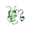
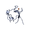
| Entry | Database: PDB / ID: 1b13 | ||||||
|---|---|---|---|---|---|---|---|
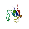
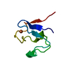
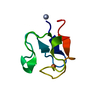
| Title | CLOSTRIDIUM PASTEURIANUM RUBREDOXIN G10A MUTANT | ||||||
 Components Components | PROTEIN (RUBREDOXIN) | ||||||
 Keywords Keywords | ELECTRON TRANSPORT / METALLOPROTEIN / IRON SULFUR / ELECTRON TRANSFER | ||||||
| Function / homology |  Function and homology information Function and homology informationalkane catabolic process / electron transfer activity / iron ion binding Similarity search - Function | ||||||
| Biological species |  Clostridium pasteurianum (bacteria) Clostridium pasteurianum (bacteria) | ||||||
| Method |  X-RAY DIFFRACTION / OTHER / Resolution: 1.5 Å X-RAY DIFFRACTION / OTHER / Resolution: 1.5 Å | ||||||
 Authors Authors | Maher, M.J. / Guss, J.M. / Wilce, M.C.J. / Wedd, A.G. | ||||||
 Citation Citation | Journal: Acta Crystallogr.,Sect.D / Year: 1999 Title: Rubredoxin from Clostridium pasteurianum. Structures of G10A, G43A and G10VG43A mutant proteins. Mutation of conserved glycine 10 to valine causes the 9-10 peptide link to invert. Authors: Maher, M.J. / Xiao, Z. / Wilce, M.C. / Guss, J.M. / Wedd, A.G. | ||||||
| History |
|
- Structure visualization
| Structure viewer | Molecule: MolmilJmol/JSmol |
|---|
- Downloads & links
-Download
| PDBx/mmCIF format | 1b13.cif.gz | 22.9 KB | Display | PDBx/mmCIF format |
|---|---|---|---|---|
| PDB format | pdb1b13.ent.gz | 13.8 KB | Display | PDB format |
| PDBx/mmJSON format | 1b13.json.gz | Tree view | PDBx/mmJSON format | |
| Others |  Other downloads Other downloads |
-Validation report
| Arichive directory | https://data.pdbj.org/pub/pdb/validation_reports/b1/1b13ftp://data.pdbj.org/pub/pdb/validation_reports/b1/1b13 | HTTPS FTP |
|---|
-Related structure data
| Related structure data |  1b2jC  1b2oC  5rxnS S: Starting model for refinement C: citing same article ( |
|---|---|
| Similar structure data |


-Links
 PDBj
PDBj
- Assembly
| Deposited unit | 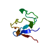
| ||||||||
|---|---|---|---|---|---|---|---|---|---|
| 1 |
| ||||||||
| Unit cell |
|
-Components
| #1: Protein | Mass: 6065.637 Da / Num. of mol.: 1 / Mutation: G10A Source method: isolated from a genetically manipulated source Source: (gene. exp.) Clostridium pasteurianum (bacteria) / Strain: JM109 / Cellular location: CYTOPLASM / Gene: CLORUB / Plasmid: PKK223-3 / Gene (production host): CLORUB / Production host: |
|---|---|
| #2: Chemical | ChemComp-FE /   Mass: 55.845 Da / Num. of mol.: 1 / Source method: obtained synthetically / Formula: Fe Mass: 55.845 Da / Num. of mol.: 1 / Source method: obtained synthetically / Formula: Fe |
| #3: Water | ChemComp-HOH /  Mass: 18.015 Da / Num. of mol.: 40 / Source method: isolated from a natural source / Formula: H2O Mass: 18.015 Da / Num. of mol.: 40 / Source method: isolated from a natural source / Formula: H2O |
-Experimental details
-Experiment
| Experiment | Method: X-RAY DIFFRACTION / Number of used crystals: 1 |
|---|
- Sample preparation
| Crystal | Density Matthews: 2.44 Å3/Da / Density % sol: 49.1 % | ||||||||||||||||||||||||||||||
|---|---|---|---|---|---|---|---|---|---|---|---|---|---|---|---|---|---|---|---|---|---|---|---|---|---|---|---|---|---|---|---|
| Crystal grow | pH: 4.6 Details: PROTEIN WAS CRYSTALLISED FROM 50-60% SATURATED AMMONIUM SULFATE IN SODIUM ACETATE BUFFER (50 MM) AT PH 4.6. | ||||||||||||||||||||||||||||||
| Components of the solutions |
| ||||||||||||||||||||||||||||||
| Crystal | *PLUS | ||||||||||||||||||||||||||||||
| Crystal grow | *PLUS Temperature: 277 K / Method: vapor diffusion, hanging drop / PH range low: 5 / PH range high: 4 | ||||||||||||||||||||||||||||||
| Components of the solutions | *PLUS
|
-Data collection
| Diffraction | Mean temperature: 293 K |
|---|---|
| Diffraction source | Source: ROTATING ANODE / Type: RIGAKU RU200 / Wavelength: 1.5418 |
| Detector | Type: RIGAKU RU200 / Detector: IMAGE PLATE / Date: Jul 1, 1996 / Details: MIRRORS |
| Radiation | Monochromator: NI FILTER 0.00015" / Protocol: SINGLE WAVELENGTH / Monochromatic (M) / Laue (L): M / Scattering type: x-ray |
| Radiation wavelength | Wavelength: 1.5418 Å / Relative weight: 1 |
| Reflection | Resolution: 1.5→30 Å / Num. obs: 6061 / % possible obs: 94.5 % / Redundancy: 3.3 % / Biso Wilson estimate: 15.74 Å2 / Rmerge(I) obs: 0.08 / Net I/σ(I): 12.5 |
| Reflection shell | Resolution: 1.5→1.55 Å / Redundancy: 1.5 % / Rmerge(I) obs: 0.245 / Mean I/σ(I) obs: 2.2 / % possible all: 58.8 |
| Reflection | *PLUS Num. obs: 7662 / Num. measured all: 29561 |
| Reflection shell | *PLUS % possible obs: 59 % |
- Processing
| Software |
| ||||||||||||||||||||||||||||||||||||||||||||||||||||||||||||||||||||||||||||||||||||
|---|---|---|---|---|---|---|---|---|---|---|---|---|---|---|---|---|---|---|---|---|---|---|---|---|---|---|---|---|---|---|---|---|---|---|---|---|---|---|---|---|---|---|---|---|---|---|---|---|---|---|---|---|---|---|---|---|---|---|---|---|---|---|---|---|---|---|---|---|---|---|---|---|---|---|---|---|---|---|---|---|---|---|---|---|---|
| Refinement | Method to determine structure: OTHER Starting model: 5RXN Resolution: 1.5→30 Å / σ(F): 0 / Details: ESD FROM CRUIKSHANK (A): 0.08
| ||||||||||||||||||||||||||||||||||||||||||||||||||||||||||||||||||||||||||||||||||||
| Displacement parameters | Biso mean: 19 Å2 | ||||||||||||||||||||||||||||||||||||||||||||||||||||||||||||||||||||||||||||||||||||
| Refinement step | Cycle: LAST / Resolution: 1.5→30 Å
| ||||||||||||||||||||||||||||||||||||||||||||||||||||||||||||||||||||||||||||||||||||
| Refine LS restraints |
| ||||||||||||||||||||||||||||||||||||||||||||||||||||||||||||||||||||||||||||||||||||
| Software | *PLUS Name: REFMAC / Classification: refinement | ||||||||||||||||||||||||||||||||||||||||||||||||||||||||||||||||||||||||||||||||||||
| Refinement | *PLUS Highest resolution: 1.5 Å / Rfactor obs: 0.171 | ||||||||||||||||||||||||||||||||||||||||||||||||||||||||||||||||||||||||||||||||||||
| Solvent computation | *PLUS | ||||||||||||||||||||||||||||||||||||||||||||||||||||||||||||||||||||||||||||||||||||
| Displacement parameters | *PLUS Biso mean: 19 Å2 | ||||||||||||||||||||||||||||||||||||||||||||||||||||||||||||||||||||||||||||||||||||
| Refine LS restraints | *PLUS
| ||||||||||||||||||||||||||||||||||||||||||||||||||||||||||||||||||||||||||||||||||||
| LS refinement shell | *PLUS Rfactor Rfree: 0.432 / Rfactor obs: 0.389 |
