 Movie
Movie Controller

[English] 日本語
 Yorodumi
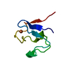
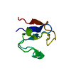
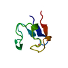
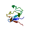
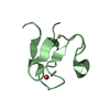
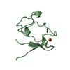
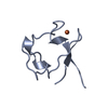
- PDB-4rxn: CRYSTALLOGRAPHIC REFINEMENT OF RUBREDOXIN AT 1.2 ANGSTROMS RESOLUTION -
+ Open data

- Basic information
| Entry | Database: PDB / ID: 4rxn | |||||||||
|---|---|---|---|---|---|---|---|---|---|---|
| Title | CRYSTALLOGRAPHIC REFINEMENT OF RUBREDOXIN AT 1.2 ANGSTROMS RESOLUTION | |||||||||
 Components Components | RUBREDOXIN | |||||||||
 Keywords Keywords | ELECTRON TRANSFER(IRON-SULFUR PROTEIN) | |||||||||
| Function / homology |  Function and homology information Function and homology informationalkane catabolic process / electron transfer activity / iron ion binding Similarity search - Function | |||||||||
| Biological species |  Clostridium pasteurianum (bacteria) Clostridium pasteurianum (bacteria) | |||||||||
| Method |  X-RAY DIFFRACTION / Resolution: 1.2 Å X-RAY DIFFRACTION / Resolution: 1.2 Å | |||||||||
 Authors Authors | Watenpaugh, K.D. / Sieker, L.C. / Jensen, L.H. | |||||||||
 Citation Citation | Journal: J.Mol.Biol. / Year: 1980 Title: Crystallographic refinement of rubredoxin at 1 x 2 A degrees resolution. Authors: Watenpaugh, K.D. / Sieker, L.C. / Jensen, L.H. #1: Journal: J.Mol.Biol. / Year: 1979Title: The Structure of Rubredoxin at 1.2 Angstroms Resolution Authors: Watenpaugh, K.D. / Sieker, L.C. / Jensen, L.H. #2: Journal: J.Mol.Biol. / Year: 1978Title: Water Structure in a Protein Crystal. Rubredoxin at 1.2 Angstroms Resolution. Authors: Watenpaugh, K.D. / Margulis, T.N. / Sieker, L.C. / Jensen, L.H. #3: Journal: Acta Crystallogr.,Sect.B / Year: 1973Title: Refinement of the Model of a Protein. Rubredoxin at 1.5 Angstroms Resolution Authors: Watenpaugh, K.D. / Sieker, L.C. / Herriott, J.R. / Jensen, L.H. #4: Journal: J.Mol.Biol. / Year: 1973Title: Sequence of Rubredoxin by X-Ray Diffraction Authors: Herriott, J.R. / Watenpaugh, K.D. / Sieker, L.C. / Jensen, L.H. #5: Journal: Cold Spring Harbor Symp.Quant.Biol. / Year: 1972Title: The Structure of a Non-Heme Iron Protein, Rubredoxin at 1.5 Angstroms Resolution Authors: Watenpaugh, K.D. / Sieker, L.C. / Herriott, J.R. / Jensen, L.H. #6: Journal: Thesis / Year: 1972Title: The Primary Structure of Clostridium Pasteurianum Rubredoxin Authors: Mccarthy, K.F. #7: Journal: J.Mol.Biol. / Year: 1970Title: Structure of Rubredoxin. An X-Ray Study to 2.5 Angstroms Resolution Authors: Herriott, J.R. / Sieker, L.C. / Jensen, L.H. / Lovenberg, W. | |||||||||
| History |
|
- Structure visualization
| Structure viewer | Molecule: MolmilJmol/JSmol |
|---|
- Downloads & links
-Download
| PDBx/mmCIF format | 4rxn.cif.gz | 34.9 KB | Display | PDBx/mmCIF format |
|---|---|---|---|---|
| PDB format | pdb4rxn.ent.gz | 26.3 KB | Display | PDB format |
| PDBx/mmJSON format | 4rxn.json.gz | Tree view | PDBx/mmJSON format | |
| Others |  Other downloads Other downloads |
-Validation report
| Arichive directory | https://data.pdbj.org/pub/pdb/validation_reports/rx/4rxnftp://data.pdbj.org/pub/pdb/validation_reports/rx/4rxn | HTTPS FTP |
|---|
-Related structure data
| Similar structure data |
|---|


-Links
 PDBj
PDBj
- Assembly
| Deposited unit | 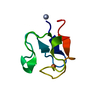
| ||||||||
|---|---|---|---|---|---|---|---|---|---|
| 1 |
| ||||||||
| Unit cell |
|
-Components
| #1: Protein | Mass: 6054.566 Da / Num. of mol.: 1 Source method: isolated from a genetically manipulated source Source: (gene. exp.) Clostridium pasteurianum (bacteria) / References: UniProt: P00268 |
|---|---|
| #2: Chemical | ChemComp-FE /   Mass: 55.845 Da / Num. of mol.: 1 / Source method: obtained synthetically / Formula: Fe Mass: 55.845 Da / Num. of mol.: 1 / Source method: obtained synthetically / Formula: Fe |
| #3: Water | ChemComp-HOH /  Mass: 18.015 Da / Num. of mol.: 127 / Source method: isolated from a natural source / Formula: H2O Mass: 18.015 Da / Num. of mol.: 127 / Source method: isolated from a natural source / Formula: H2O |
-Experimental details
-Experiment
| Experiment | Method: X-RAY DIFFRACTION |
|---|
- Sample preparation
| Crystal | Density Matthews: 2.13 Å3/Da / Density % sol: 42.32 % |
|---|---|
| Crystal grow | *PLUS Temperature: 23 ℃ / pH: 4 / Method: unknown / Details: salting out |
| Components of the solutions | *PLUS Conc.: 80 %sat / Chemical formula: (NH4)2SO4 |
-Data collection
| Reflection | *PLUS Observed criterion σ(I): 4 / Num. measured all: 17000 / Rmerge(I) obs: 0.038 |
|---|
- Processing
| Refinement | Resolution: 1.2→10 Å Details: EACH HYDROGEN ATOM POSITION HAS BEEN CALCULATED FROM THE COORDINATES OF THE ATOM IT IS BONDED TO. THE B-VALUE OF THE HEAVIER ATOM WAS ASSIGNED TO THE HYDROGEN ATOM. THE CELL DIMENSIONS ...Details: EACH HYDROGEN ATOM POSITION HAS BEEN CALCULATED FROM THE COORDINATES OF THE ATOM IT IS BONDED TO. THE B-VALUE OF THE HEAVIER ATOM WAS ASSIGNED TO THE HYDROGEN ATOM. THE CELL DIMENSIONS VARIED FROM START TO FINISH OF DATA COLLECTION. THE VALUES GIVEN ARE THOSE AT THE MID-POINT OF DATA COLLECTION.
| ||||||||||||
|---|---|---|---|---|---|---|---|---|---|---|---|---|---|
| Refinement step | Cycle: LAST / Resolution: 1.2→10 Å
| ||||||||||||
| Refinement | *PLUS Highest resolution: 1.2 Å / Lowest resolution: 10 Å / Num. reflection obs: 10936 / σ(I): 2 / Rfactor obs: 0.128 | ||||||||||||
| Solvent computation | *PLUS | ||||||||||||
| Displacement parameters | *PLUS Biso mean: 12 Å2 |
