 Movie
Movie Controller

[English] 日本語
 Yorodumi
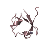
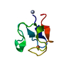
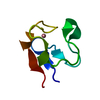
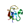
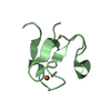
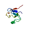
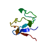
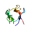
- PDB-1smw: Crystal Structure of Cp Rd L41A mutant in reduced state 2 (soaked) -
+ Open data

- Basic information
| Entry | Database: PDB / ID: 1smw | ||||||
|---|---|---|---|---|---|---|---|
| Title | Crystal Structure of Cp Rd L41A mutant in reduced state 2 (soaked) | ||||||
 Components Components | Rubredoxin | ||||||
 Keywords Keywords | ELECTRON TRANSPORT | ||||||
| Function / homology |  Function and homology information Function and homology informationalkane catabolic process / electron transfer activity / iron ion binding Similarity search - Function | ||||||
| Biological species |  Clostridium pasteurianum (bacteria) Clostridium pasteurianum (bacteria) | ||||||
| Method |  X-RAY DIFFRACTION / SYNCHROTRON / MOLECULAR REPLACEMENT / Resolution: 1.38 Å X-RAY DIFFRACTION / SYNCHROTRON / MOLECULAR REPLACEMENT / Resolution: 1.38 Å | ||||||
 Authors Authors | Park, I.Y. / Youn, B. / Harley, J.L. / Eidsness, M.K. / Smith, E. / Ichiye, T. / Kang, C. | ||||||
 Citation Citation | Journal: J.BIOL.INORG.CHEM. / Year: 2004 Title: The unique hydrogen bonded water in the reduced form of Clostridium pasteurianum rubredoxin and its possible role in electron transfer Authors: Park, I.Y. / Youn, B. / Harley, J.L. / Eidsness, M.K. / Smith, E. / Ichiye, T. / Kang, C. | ||||||
| History |
|
- Structure visualization
| Structure viewer | Molecule: MolmilJmol/JSmol |
|---|
- Downloads & links
-Download
| PDBx/mmCIF format | 1smw.cif.gz | 31.1 KB | Display | PDBx/mmCIF format |
|---|---|---|---|---|
| PDB format | pdb1smw.ent.gz | 21 KB | Display | PDB format |
| PDBx/mmJSON format | 1smw.json.gz | Tree view | PDBx/mmJSON format | |
| Others |  Other downloads Other downloads |
-Validation report
| Arichive directory | https://data.pdbj.org/pub/pdb/validation_reports/sm/1smwftp://data.pdbj.org/pub/pdb/validation_reports/sm/1smw | HTTPS FTP |
|---|
-Related structure data
| Related structure data |  1smmC  1smuC  1fhhS C: citing same article ( S: Starting model for refinement |
|---|---|
| Similar structure data |

-Links
 PDBj
PDBj


- Assembly
| Deposited unit | 
| ||||||||
|---|---|---|---|---|---|---|---|---|---|
| 1 |
| ||||||||
| Unit cell |
|
-Components
| #1: Protein | Mass: 6009.531 Da / Num. of mol.: 1 / Mutation: L41A Source method: isolated from a genetically manipulated source Source: (gene. exp.) Clostridium pasteurianum (bacteria) / Production host: |
|---|---|
| #2: Chemical | ChemComp-FE2 /   Mass: 55.845 Da / Num. of mol.: 1 / Source method: obtained synthetically / Formula: Fe Mass: 55.845 Da / Num. of mol.: 1 / Source method: obtained synthetically / Formula: Fe |
| #3: Water | ChemComp-HOH /  Mass: 18.015 Da / Num. of mol.: 48 / Source method: isolated from a natural source / Formula: H2O Mass: 18.015 Da / Num. of mol.: 48 / Source method: isolated from a natural source / Formula: H2O |
-Experimental details
-Experiment
| Experiment | Method: X-RAY DIFFRACTION / Number of used crystals: 1 |
|---|
- Sample preparation
| Crystal | Density Matthews: 2.08 Å3/Da / Density % sol: 40.83 % | ||||||||||||||||||||||||
|---|---|---|---|---|---|---|---|---|---|---|---|---|---|---|---|---|---|---|---|---|---|---|---|---|---|
| Crystal grow | Temperature: 277 K / Method: vapor diffusion, hanging drop / pH: 4 Details: ammonium sulfate, sodium chloride, sodium dithionite, pH 4.0, VAPOR DIFFUSION, HANGING DROP, temperature 277K | ||||||||||||||||||||||||
| Crystal grow | *PLUS Method: vapor diffusion, hanging drop | ||||||||||||||||||||||||
| Components of the solutions | *PLUS
|
-Data collection
| Diffraction | Mean temperature: 113 K |
|---|---|
| Diffraction source | Source: SYNCHROTRON / Site: ALS  / Beamline: 8.2.1 / Wavelength: 1.0332 Å / Beamline: 8.2.1 / Wavelength: 1.0332 Å |
| Detector | Type: ADSC QUANTUM 210 / Detector: CCD / Date: Jul 31, 2003 |
| Radiation | Protocol: SINGLE WAVELENGTH / Monochromatic (M) / Laue (L): M / Scattering type: x-ray |
| Radiation wavelength | Wavelength: 1.0332 Å / Relative weight: 1 |
| Reflection | Resolution: 1.38→20 Å / Num. all: 9923 / Num. obs: 9923 / % possible obs: 99.3 % / Observed criterion σ(F): 2 / Observed criterion σ(I): 1.4 |
| Reflection shell | Resolution: 1.38→1.47 Å / % possible all: 97.8 |
| Reflection | *PLUS Rmerge(I) obs: 0.038 |
| Reflection shell | *PLUS Rmerge(I) obs: 0.125 / Mean I/σ(I) obs: 2.5 |
- Processing
| Software |
| ||||||||||||||||||||
|---|---|---|---|---|---|---|---|---|---|---|---|---|---|---|---|---|---|---|---|---|---|
| Refinement | Method to determine structure: MOLECULAR REPLACEMENT Starting model: PDB ENTRY 1FHH Resolution: 1.38→20 Å / Cross valid method: THROUGHOUT / σ(F): 0.001 / Stereochemistry target values: Engh & Huber
| ||||||||||||||||||||
| Refinement step | Cycle: LAST / Resolution: 1.38→20 Å
| ||||||||||||||||||||
| Refinement | *PLUS Rfactor Rfree: 0.2 | ||||||||||||||||||||
| Solvent computation | *PLUS | ||||||||||||||||||||
| Displacement parameters | *PLUS | ||||||||||||||||||||
| Refine LS restraints | *PLUS
|
