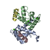
COPI-independent Golgi-to-ER retrograde traffic / Advanced glycosylation endproduct receptor signaling / Factors involved in megakaryocyte development and platelet production / Regulation of TLR by endogenous ligand / HSP90 chaperone cycle for steroid hormone receptors (SHR) in the presence of ligand / COPI-mediated anterograde transport / F-actin capping protein complex / MHC class II antigen presentation / A band / barbed-end actin filament capping ...COPI-independent Golgi-to-ER retrograde traffic / Advanced glycosylation endproduct receptor signaling / Factors involved in megakaryocyte development and platelet production / Regulation of TLR by endogenous ligand / HSP90 chaperone cycle for steroid hormone receptors (SHR) in the presence of ligand / COPI-mediated anterograde transport / F-actin capping protein complex / MHC class II antigen presentation / A band / barbed-end actin filament capping / S100 protein binding / M band / I band / positive regulation of sprouting angiogenesis / regulation of heart contraction / cortical cytoskeleton / brush border / sarcoplasmic reticulum / Z disc / calcium-dependent protein binding / actin filament binding / ATPase binding / actin cytoskeleton organization / calcium ion binding / negative regulation of transcription by RNA polymerase II / Golgi apparatus / protein homodimerization activity / mitochondrion / nucleoplasm / identical protein binding / nucleus / membrane / cytosol / cytoplasm Similarity search - Function
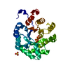
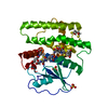
Protein S100-A1 / F-actin capping protein, alpha subunit, conserved site / F-actin capping protein alpha subunit signature 1. / F-actin capping protein alpha subunit signature 2. / F-actin-capping protein subunit alpha / F-actin-capping protein subunit alpha/beta / F-actin-capping protein subunit alpha/beta, domain 2 / F-actin capping protein, alpha subunit, domain 1 / F-actin capping protein alpha subunit / S-100/ICaBP type calcium binding protein signature. ...Protein S100-A1 / F-actin capping protein, alpha subunit, conserved site / F-actin capping protein alpha subunit signature 1. / F-actin capping protein alpha subunit signature 2. / F-actin-capping protein subunit alpha / F-actin-capping protein subunit alpha/beta / F-actin-capping protein subunit alpha/beta, domain 2 / F-actin capping protein, alpha subunit, domain 1 / F-actin capping protein alpha subunit / S-100/ICaBP type calcium binding protein signature. / S100/Calcium binding protein 7/8-like, conserved site / S100/CaBP-9k-type, calcium binding, subdomain / S-100/ICaBP type calcium binding domain / S-100/ICaBP type calcium binding domain / EF hand domain / EF-hand / Recoverin; domain 1 / EF-hand, calcium binding motif / EF-Hand 1, calcium-binding site / EF-hand calcium-binding domain. / EF-hand calcium-binding domain profile. / EF-hand domain / EF-hand domain pair / Orthogonal Bundle / Mainly Alpha Similarity search - Domain/homology
Conformer selection criteria: structures with the least restraint violations Conformers calculated total number: 200 / Conformers submitted total number: 20
+
About Yorodumi
-
News
-
Feb 9, 2022. New format data for meta-information of EMDB entries
New format data for meta-information of EMDB entries
Version 3 of the EMDB header file is now the official format.
The previous official version 1.9 will be removed from the archive.
In the structure databanks used in Yorodumi, some data are registered as the other names, "COVID-19 virus" and "2019-nCoV". Here are the details of the virus and the list of structure data.
Jan 31, 2019. EMDB accession codes are about to change! (news from PDBe EMDB page)
EMDB accession codes are about to change! (news from PDBe EMDB page)
The allocation of 4 digits for EMDB accession codes will soon come to an end. Whilst these codes will remain in use, new EMDB accession codes will include an additional digit and will expand incrementally as the available range of codes is exhausted. The current 4-digit format prefixed with “EMD-” (i.e. EMD-XXXX) will advance to a 5-digit format (i.e. EMD-XXXXX), and so on. It is currently estimated that the 4-digit codes will be depleted around Spring 2019, at which point the 5-digit format will come into force.
The EM Navigator/Yorodumi systems omit the EMD- prefix.
Related info.:Q: What is EMD? / ID/Accession-code notation in Yorodumi/EM Navigator
Yorodumi is a browser for structure data from EMDB, PDB, SASBDB, etc.
This page is also the successor to EM Navigator detail page, and also detail information page/front-end page for Omokage search.
The word "yorodu" (or yorozu) is an old Japanese word meaning "ten thousand". "mi" (miru) is to see.
Related info.:EMDB / PDB / SASBDB / Comparison of 3 databanks / Yorodumi Search / Aug 31, 2016. New EM Navigator & Yorodumi / Yorodumi Papers / Jmol/JSmol / Function and homology information / Changes in new EM Navigator and Yorodumi
 Movie
Movie Controller


 Open data
 Basic information
 Components
 Keywords
 Function and homology information
 Authors
 Citation
 Structure visualization
 Downloads & links
 Other downloads


 PDBj
PDBj


 Assembly


 Mass: 40.078 Da / Num. of mol.: 4 / Source method: obtained synthetically / Formula: Ca
 HSQC
HSQC Sample preparation
 Processing