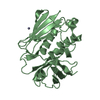
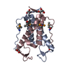
- PDB-1zfs: Solution structure of S100A1 bound to calcium -
+
Open data
ID or keywords:
Loading...
-
Basic information
Entry
Database: PDB / ID: 1zfs
Title
Solution structure of S100A1 bound to calcium
Components
S-100 protein, alpha chain
Keywords
METAL BINDING PROTEIN / S100 / noncovalent homodimer / x-type 4 helix bundle / calcium binding / conformational change
Function / homology
Function and homology information
Regulation of TLR by endogenous ligand / A band / S100 protein binding / M band / I band / positive regulation of sprouting angiogenesis / regulation of heart contraction / sarcoplasmic reticulum / vasodilation / Z disc ...Regulation of TLR by endogenous ligand / A band / S100 protein binding / M band / I band / positive regulation of sprouting angiogenesis / regulation of heart contraction / sarcoplasmic reticulum / vasodilation / Z disc / calcium-dependent protein binding / ATPase binding / calcium ion binding / negative regulation of transcription by RNA polymerase II / Golgi apparatus / protein homodimerization activity / mitochondrion / nucleoplasm / identical protein binding / nucleus / cytoplasm / cytosol Similarity search - Function
Protein S100-A1 / S-100/ICaBP type calcium binding protein signature. / S100/Calcium binding protein 7/8-like, conserved site / S100/CaBP-9k-type, calcium binding, subdomain / S-100/ICaBP type calcium binding domain / S-100/ICaBP type calcium binding domain / EF hand domain / EF-hand / Recoverin; domain 1 / EF-hand, calcium binding motif ...Protein S100-A1 / S-100/ICaBP type calcium binding protein signature. / S100/Calcium binding protein 7/8-like, conserved site / S100/CaBP-9k-type, calcium binding, subdomain / S-100/ICaBP type calcium binding domain / S-100/ICaBP type calcium binding domain / EF hand domain / EF-hand / Recoverin; domain 1 / EF-hand, calcium binding motif / EF-Hand 1, calcium-binding site / EF-hand calcium-binding domain. / EF-hand calcium-binding domain profile. / EF-hand domain / EF-hand domain pair / Orthogonal Bundle / Mainly Alpha Similarity search - Domain/homology
Mass: 40.078 Da / Num. of mol.: 4 / Source method: obtained synthetically / Formula: Ca
-
Experimental details
-
Experiment
Experiment
Method: SOLUTION NMR
NMR experiment
Conditions-ID
Experiment-ID
Solution-ID
Type
1
1
1
4D 13C-separated NOESY
1
2
1
4D 13C/15N-separated NOESY
1
3
1
3D 15N-separated NOESY
1
4
1
HNHA
NMR details
Text: RDC data using radially and axially compressed acrylamide gels were added into structure calculations. H-bonds were determined using hydrogen exchange experiments
-
Sample preparation
Details
Contents: 0.5-3 mM S100A1 monomer, U-15N, 13C, 0.35 mM NaN3, 15 mM NaCl, 20 mM CaCl2, 25 mM Tris, 25 mM DTT, 10% D2O Solvent system: 90% H2O/10% D2O
Sample conditions
Ionic strength: 15 mM / pH: 7.2 / Pressure: ambient / Temperature: 310 K
-
NMR measurement
Radiation
Protocol: SINGLE WAVELENGTH / Monochromatic (M) / Laue (L): M
Radiation wavelength
Relative weight: 1
NMR spectrometer
Type
Manufacturer
Model
Field strength (MHz)
Spectrometer-ID
Bruker DMX
Bruker
DMX
600
1
Bruker AVANCE
Bruker
AVANCE
800
2
-
Processing
NMR software
Name
Version
Developer
Classification
X-PLOR
2.9.3
C.D. Schwieters, J.J. Kuszewski, N. Tjandra, G.M. Clore
structuresolution
X-PLOR
2.9.3
C.D. Schwieters, J.J. Kuszewski, N. Tjandra, G.M. Clore
Conformer selection criteria: structures with the least restraint violations Conformers calculated total number: 200 / Conformers submitted total number: 19
+
About Yorodumi
-
News
-
Feb 9, 2022. New format data for meta-information of EMDB entries
New format data for meta-information of EMDB entries
Version 3 of the EMDB header file is now the official format.
The previous official version 1.9 will be removed from the archive.
In the structure databanks used in Yorodumi, some data are registered as the other names, "COVID-19 virus" and "2019-nCoV". Here are the details of the virus and the list of structure data.
Jan 31, 2019. EMDB accession codes are about to change! (news from PDBe EMDB page)
EMDB accession codes are about to change! (news from PDBe EMDB page)
The allocation of 4 digits for EMDB accession codes will soon come to an end. Whilst these codes will remain in use, new EMDB accession codes will include an additional digit and will expand incrementally as the available range of codes is exhausted. The current 4-digit format prefixed with “EMD-” (i.e. EMD-XXXX) will advance to a 5-digit format (i.e. EMD-XXXXX), and so on. It is currently estimated that the 4-digit codes will be depleted around Spring 2019, at which point the 5-digit format will come into force.
The EM Navigator/Yorodumi systems omit the EMD- prefix.
Related info.:Q: What is EMD? / ID/Accession-code notation in Yorodumi/EM Navigator
Yorodumi is a browser for structure data from EMDB, PDB, SASBDB, etc.
This page is also the successor to EM Navigator detail page, and also detail information page/front-end page for Omokage search.
The word "yorodu" (or yorozu) is an old Japanese word meaning "ten thousand". "mi" (miru) is to see.
Related info.:EMDB / PDB / SASBDB / Comparison of 3 databanks / Yorodumi Search / Aug 31, 2016. New EM Navigator & Yorodumi / Yorodumi Papers / Jmol/JSmol / Function and homology information / Changes in new EM Navigator and Yorodumi
 Movie
Movie Controller


 Open data
 Basic information
 Components
 Keywords
 Function and homology information
 Authors
 Citation
 Structure visualization
 Downloads & links
 Other downloads

 PDBj
PDBj Assembly
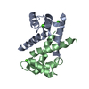


 Mass: 40.078 Da / Num. of mol.: 4 / Source method: obtained synthetically / Formula: Ca
 Sample preparation
 Processing
 X-PLOR