 Movie
Movie Controller

[English] 日本語
 Yorodumi
- PDB-1kga: STRUCTURE OF 2-KETO-3-DEOXY-6-PHOSPHOGLUCONATE ALDOLASE AT 2.8 AN... -
+ Open data

- Basic information
| Entry | Database: PDB / ID: 1kga | ||||||
|---|---|---|---|---|---|---|---|
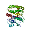
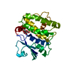
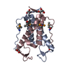
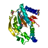
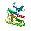
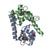
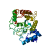
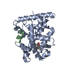
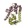
| Title | STRUCTURE OF 2-KETO-3-DEOXY-6-PHOSPHOGLUCONATE ALDOLASE AT 2.8 ANGSTROMS RESOLUTION | ||||||
 Components Components | 2-KETO-3-DEOXY-6-PHOSPHOGLUCONATE ALDOLASE | ||||||
 Keywords Keywords | LYASE / LYASE (ALDEHYDE) | ||||||
| Biological species |  Pseudomonas putida (bacteria) Pseudomonas putida (bacteria) | ||||||
| Method |  X-RAY DIFFRACTION / Resolution: 3.5 Å X-RAY DIFFRACTION / Resolution: 3.5 Å | ||||||
 Authors Authors | Tulinsky, A. | ||||||
 Citation Citation | Journal: J.Mol.Biol. / Year: 1982 Title: Structure of 2-keto-3-deoxy-6-phosphogluconate aldolase at 2 . 8 A resolution. Authors: Mavridis, I.M. / Hatada, M.H. / Tulinsky, A. / Lebioda, L. #1: Journal: J.Mol.Biol. / Year: 1982Title: Comparison of the Folding of 2-Keto-3-Deoxy-6-Phosphogluconate Aldolase, Triosephosphate Isomerase and Pyruvate Kinase. Implications in Molecular Evolution Authors: Lebioda, L. / Hatada, M.H. / Tulinsky, A. / Mavridis, I.M. #2: Journal: Am.Cryst.Assoc.,Abstr.Papers (Winter Meeting) / Year: 1978Title: The Folding and Structure of 2-Keto-3-Deoxy-6-Phosphogluconic Aldolase (Kdpg Aldolase) from Pseudomonas Putida, Interpreted in Light of the Amino Acid Sequence Authors: Frentup, M. / Weber, L.D. / Tulinsky, A. #3: Journal: Biochemistry / Year: 1976Title: The Folding and Quaternary Structure of Trimeric 2-Keto-3-Deoxy-6-Phosphogluconic Aldolase at 3.5-Angstroms Resolution Authors: Mavridis, I.M. / Tulinsky, A. #4: Journal: J.Biol.Chem. / Year: 1973Title: Confirmation of a Trimeric Subunit Arrangement for 2-Keto-3-Deoxy-6-Phosphogluconic Aldolase Using X-Ray Crystallographic Methods Authors: Vandlen, R.L. / Ersfeld, D.L. / Tulinsky, A. / Wood, W.A. | ||||||
| History |
|
- Structure visualization
| Structure viewer | Molecule:  MolmilJmol/JSmol MolmilJmol/JSmol |
|---|
- Downloads & links
-Download
| PDBx/mmCIF format | 1kga.cif.gz | 11.1 KB | Display | PDBx/mmCIF format |
|---|---|---|---|---|
| PDB format | pdb1kga.ent.gz | 7 KB | Display | PDB format |
| PDBx/mmJSON format | 1kga.json.gz | Tree view | PDBx/mmJSON format | |
| Others |  Other downloads Other downloads |
-Validation report
| Arichive directory | https://data.pdbj.org/pub/pdb/validation_reports/kg/1kgaftp://data.pdbj.org/pub/pdb/validation_reports/kg/1kga | HTTPS FTP |
|---|
-Related structure data
| Similar structure data |
|---|

-Links
 PDBj
PDBj- Assembly
| Deposited unit | 
| ||||||||||||
|---|---|---|---|---|---|---|---|---|---|---|---|---|---|
| 1 |
| ||||||||||||
| Unit cell |
| ||||||||||||
| Noncrystallographic symmetry (NCS) | NCS oper:
|
-Components
| #1: Protein | Mass: 18655.930 Da / Num. of mol.: 1 Source method: isolated from a genetically manipulated source Source: (gene. exp.) Pseudomonas putida (bacteria) / References: 2-dehydro-3-deoxy-phosphogluconate aldolase |
|---|---|
| Sequence details | RECENT SEQUENCE INFORMATION FROM W.A.WOOD AND CO-WORKERS IMPLIES THE PRESENCE OF ABOUT 20 AMINO ...RECENT SEQUENCE INFORMATIO |
-Experimental details
-Experiment
| Experiment | Method: X-RAY DIFFRACTION |
|---|
- Sample preparation
| Crystal | Density Matthews: 4.94 Å3/Da / Density % sol: 75.09 % |
|---|---|
| Crystal grow | *PLUS Method: other / Details: Vandlen, R.L., (1973) J. Biol. Chem., 248, 2251. |
-Data collection
| Radiation | Scattering type: x-ray |
|---|---|
| Radiation wavelength | Relative weight: 1 |
- Processing
| Refinement | Highest resolution: 3.5 Å Details: THE ORTHOGONAL AXIAL SYSTEM OF THIS ENTRY IS NOT THAT OF THE STANDARD REPRESENTATION OF THE SPACE GROUP P 21 3. THE *Z* AXIS OF THIS ENTRY IS PARALLEL TO A THREE-FOLD AXIS OF THE SPACE GROUP ...Details: THE ORTHOGONAL AXIAL SYSTEM OF THIS ENTRY IS NOT THAT OF THE STANDARD REPRESENTATION OF THE SPACE GROUP P 21 3. THE *Z* AXIS OF THIS ENTRY IS PARALLEL TO A THREE-FOLD AXIS OF THE SPACE GROUP AND CORRESPONDS TO A DIRECTION ALIGNED WITH A BODY DIAGONAL OF THE CONVENTIONAL CUBIC UNIT CELL. THIS TRIMER-FORMING AXIS PASSES THROUGH THE POINT X=53.2, Y=-45.2. THE TWO TRANSFORMATIONS GIVEN IN THE *MTRIX* RECORDS BELOW MAY BE USED TO GENERATE COORDINATES FOR THE REMAINING TWO SUBUNITS OF THE TRIMER IN THE COORDINATE SYSTEM OF THIS ENTRY. NOTE THAT THE THREE-FOLD AXIS INDICATED HERE IS A TRUE CRYSTALLOGRAPHIC SYMMETRY ELEMENT BUT IT IS EXPRESSED WITH RESPECT TO THE NON-STANDARD AXIAL SYSTEM. | ||||||||||||
|---|---|---|---|---|---|---|---|---|---|---|---|---|---|
| Refinement step | Cycle: LAST / Highest resolution: 3.5 Å
|
