 Movie
Movie Controller

[English] 日本語
 Yorodumi
- PDB-2k1g: Solution NMR structure of lipoprotein spr from Escherichia coli K... -
+ Open data

- Basic information
| Entry | Database: PDB / ID: 2k1g | |||||||||
|---|---|---|---|---|---|---|---|---|---|---|
| Title | Solution NMR structure of lipoprotein spr from Escherichia coli K12. Northeast Structural Genomics target ER541-37-162 | |||||||||
 Components Components | Lipoprotein spr | |||||||||
 Keywords Keywords | LIPOPROTEIN / solution NMR structure / bacterial lipoprotein / cysteine peptidase / NplC/P60 family / construct optimized / Membrane / Palmitate / Structural Genomics / PSI-2 / Protein Structure Initiative / Northeast Structural Genomics Consortium / NESG | |||||||||
| Function / homology |  Function and homology information Function and homology informationmuramoyltetrapeptide carboxypeptidase / muramoyltetrapeptide carboxypeptidase activity / capsule polysaccharide biosynthetic process / peptidoglycan metabolic process / peptidoglycan turnover / Hydrolases; Acting on peptide bonds (peptidases) / cysteine-type peptidase activity / cell outer membrane / cell wall organization / endopeptidase activity / proteolysis Similarity search - Function | |||||||||
| Biological species |  | |||||||||
| Method | SOLUTION NMR / simulated annealing | |||||||||
 Authors Authors | Aramini, J.M. / Rossi, P. / Zhao, L. / Jiang, M. / Maglaqui, M. / Xiao, R. / Liu, J. / Baran, M.C. / Swapna, G.V.T. / Huang, Y.J. ...Aramini, J.M. / Rossi, P. / Zhao, L. / Jiang, M. / Maglaqui, M. / Xiao, R. / Liu, J. / Baran, M.C. / Swapna, G.V.T. / Huang, Y.J. / Acton, T.B. / Rost, B. / Montelione, G.T. / Northeast Structural Genomics Consortium (NESG) | |||||||||
 Citation Citation | Journal: Biochemistry / Year: 2008 Title: Solution NMR structure of the NlpC/P60 domain of lipoprotein Spr from Escherichia coli: structural evidence for a novel cysteine peptidase catalytic triad. Authors: Aramini, J.M. / Rossi, P. / Huang, Y.J. / Zhao, L. / Jiang, M. / Maglaqui, M. / Xiao, R. / Locke, J. / Nair, R. / Rost, B. / Acton, T.B. / Inouye, M. / Montelione, G.T. | |||||||||
| History |
|
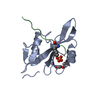
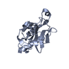
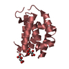
- Structure visualization
| Structure viewer | Molecule: MolmilJmol/JSmol |
|---|
- Downloads & links
-Download
| PDBx/mmCIF format | 2k1g.cif.gz | 920.7 KB | Display | PDBx/mmCIF format |
|---|---|---|---|---|
| PDB format | pdb2k1g.ent.gz | 782.1 KB | Display | PDB format |
| PDBx/mmJSON format | 2k1g.json.gz | Tree view | PDBx/mmJSON format | |
| Others |  Other downloads Other downloads |
-Validation report
| Arichive directory | https://data.pdbj.org/pub/pdb/validation_reports/k1/2k1gftp://data.pdbj.org/pub/pdb/validation_reports/k1/2k1g | HTTPS FTP |
|---|
-Related structure data
| Similar structure data | |
|---|---|
| Other databases |


-Links
 PDBj
PDBj


- Assembly
| Deposited unit | 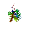
| |||||||||
|---|---|---|---|---|---|---|---|---|---|---|
| 1 |
| |||||||||
| NMR ensembles |
|
-Components
| #1: Protein | Mass: 15584.570 Da / Num. of mol.: 1 / Fragment: Residues 63-188 Source method: isolated from a genetically manipulated source Source: (gene. exp.) |
|---|
-Experimental details
-Experiment
| Experiment | Method: SOLUTION NMR | ||||||||||||||||||||||||||||||||||||||||||||||||||||||||||||||||||||||||||||
|---|---|---|---|---|---|---|---|---|---|---|---|---|---|---|---|---|---|---|---|---|---|---|---|---|---|---|---|---|---|---|---|---|---|---|---|---|---|---|---|---|---|---|---|---|---|---|---|---|---|---|---|---|---|---|---|---|---|---|---|---|---|---|---|---|---|---|---|---|---|---|---|---|---|---|---|---|---|
| NMR experiment |
| ||||||||||||||||||||||||||||||||||||||||||||||||||||||||||||||||||||||||||||
| NMR details | Text: THE PROTEIN IS MONOMERIC BY GEL FILTRATION CHROMATOGRAPHY AND STATIC LIGHT SCATTERING. THE STRUCTURE WAS DETERMINED USING TRIPLE RESONANCE NMR SPECTROSCOPY. AUTOMATED BACKBONE ASSIGNMENTS WERE ...Text: THE PROTEIN IS MONOMERIC BY GEL FILTRATION CHROMATOGRAPHY AND STATIC LIGHT SCATTERING. THE STRUCTURE WAS DETERMINED USING TRIPLE RESONANCE NMR SPECTROSCOPY. AUTOMATED BACKBONE ASSIGNMENTS WERE MADE USING AUTOASSIGN, AND THE SIDE CHAIN ASSIGNMENTS WERE COMPLETED MANUALLY. AUTOMATIC NOESY ASSIGNMENTS WERE DETERMINED USING CYANA 2.1. DIHEDRAL ANGLE CONSTRAINTS WERE OBTAINED FROM TALOS. HYDROGEN BOND CONSTRAINTS WERE DETERMINED USING BOTH AUTOSTRUCTURE AND CYANA, AND WERE APPLIED ONLY IN THE FINAL REFINEMENT STAGE (CNS) OF THE STRUCTURE DETERMINATION. COMPLETENESS OF NMR ASSIGNMENTS (EXCLUDING C-TERMINAL HHHHHH): BACKBONE, 97.9%, SIDE CHAIN, 94.2%, AROMATICS, 91.8%, STEREOSPECIFIC METHYL, 100%, STEREOSPECIFIC SIDE CHAIN NH2: 83.3%. FINAL STRUCTURE QUALITY FACTORS (FOR RESIDUES 37 TO 162, PSVS 1.3), WHERE ORDERED RESIDUES [S(PHI) + S(PSI) > 1.8] COMPRISE: 38-58,61-93,96-113,118-124,128-159: (A) RMSD (ORDERED RESIDUES): BB, 0.5, HEAVY ATOM, 1.0. (B) RAMACHANDRAN STATISTICS FOR ORDERED RESIDUES: MOST FAVORED, 88.4%, ADDITIONALLY ALLOWED, 11.5%, GENEROUSLY ALLOWED, 0.1%, DISALLOWED, 0.0%. (C) PROCHECK SCORES FOR ORDERED RESIDUES (RAW/Z-): PHI-PSI, -0.46/-1.49, ALL, -0.27/-1.60. (D) MOLPROBITY CLASH SCORE (RAW/Z-): 18.98/-1.73. (E) RPF SCORES FOR GOODNESS OF FIT TO NOESY DATA (RESIDUES 37-162): RECALL, 0.985, PRECISION, 0.925, F-MEASURE, 0.954, DP-SCORE, 0.816. (F) NUMBER OF CLOSE CONTACTS PER 20 MODELS: 8. THE C-TERMINAL HIS TAG RESIDUES OF THE PROTEIN (HHHHHH) WERE NOT INCLUDED IN THE STRUCTURE CALCULATIONS AND HAVE BEEN OMITTED FROM THIS DEPOSITION. COORDINATES FOR THE FOLLOWING RESIDUES ARE NOT WELL DETERMINED [S(PHI) + S(PSI) < 1.8]: 37,59-60,94-95,114-117,125-127,160-162. |
 HSQC
HSQC- Sample preparation
| Details |
| ||||||||||||||||||||||||||||||||||||||||||||||||||||||||||||||||||||||||||||
|---|---|---|---|---|---|---|---|---|---|---|---|---|---|---|---|---|---|---|---|---|---|---|---|---|---|---|---|---|---|---|---|---|---|---|---|---|---|---|---|---|---|---|---|---|---|---|---|---|---|---|---|---|---|---|---|---|---|---|---|---|---|---|---|---|---|---|---|---|---|---|---|---|---|---|---|---|---|
| Sample |
| ||||||||||||||||||||||||||||||||||||||||||||||||||||||||||||||||||||||||||||
| Sample conditions | Ionic strength: 0.1 / pH: 6.5 / Pressure: ambient / Temperature: 298 K |
-NMR measurement
| NMR spectrometer |
|
|---|
- Processing
| NMR software |
| ||||||||||||||||||||||||||||||||||||||||||||||||||||
|---|---|---|---|---|---|---|---|---|---|---|---|---|---|---|---|---|---|---|---|---|---|---|---|---|---|---|---|---|---|---|---|---|---|---|---|---|---|---|---|---|---|---|---|---|---|---|---|---|---|---|---|---|---|
| Refinement | Method: simulated annealing / Software ordinal: 1 Details: THE FINAL STRUCTURES ARE BASED ON A TOTAL OF 2525 CONFORMATIONALLY-RESTRICTING NOE-DERIVED DISTANCE CONSTRAINTS, 138 DIHEDRAL ANGLE CONSTRAINTS, AND 48 HYDROGEN BOND CONSTRAINTS (21.0 ...Details: THE FINAL STRUCTURES ARE BASED ON A TOTAL OF 2525 CONFORMATIONALLY-RESTRICTING NOE-DERIVED DISTANCE CONSTRAINTS, 138 DIHEDRAL ANGLE CONSTRAINTS, AND 48 HYDROGEN BOND CONSTRAINTS (21.0 CONSTRAINTS PER RESIDUE, 7.1 LONG RANGE CONSTRAINTS PER RESIDUE, COMPUTED FOR RESIDUES 37 TO 162 BY PSVS 1.3). STRUCTURE DETERMINATION WAS PERFORMED ITERATIVELY USING CYANA 2.1. THE 20 STRUCTURES OUT OF 100 WITH THE LOWEST TARGET FUNCTION WERE FURTHER REFINED BY RESTRAINED MOLECULAR DYNAMICS/ENERGY MINIMIZATION IN EXPLICIT WATER (CNS) WITH PARAM19, AND USING NEUTRAL HISTIDINES AT POSITIONS 119 and 131. | ||||||||||||||||||||||||||||||||||||||||||||||||||||
| NMR constraints | NOE constraints total: 2525 / NOE intraresidue total count: 696 / NOE long range total count: 890 / NOE medium range total count: 383 / NOE sequential total count: 556 / Hydrogen bond constraints total count: 48 / Protein phi angle constraints total count: 69 / Protein psi angle constraints total count: 69 | ||||||||||||||||||||||||||||||||||||||||||||||||||||
| NMR representative | Selection criteria: lowest energy | ||||||||||||||||||||||||||||||||||||||||||||||||||||
| NMR ensemble | Average torsion angle constraint violation: 0.2 ° Conformer selection criteria: structures with the lowest energy Conformers calculated total number: 100 / Conformers submitted total number: 20 / Maximum torsion angle constraint violation: 2.7 ° / Maximum upper distance constraint violation: 0.41 Å / Torsion angle constraint violation method: PSVS | ||||||||||||||||||||||||||||||||||||||||||||||||||||
| NMR ensemble rms | Distance rms dev: 0.01 Å |
