 Movie
Movie Controller

[English] 日本語
 Yorodumi
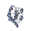
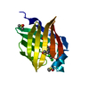
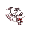
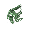
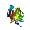
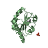
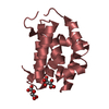
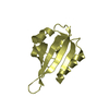
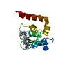
- PDB-3i86: Crystal structure of the P60 Domain from M. avium subspecies para... -
+ Open data

- Basic information
| Entry | Database: PDB / ID: 3i86 | ||||||
|---|---|---|---|---|---|---|---|
| Title | Crystal structure of the P60 Domain from M. avium subspecies paratuberculosis antigen MAP1204 | ||||||
 Components Components | Putative uncharacterized protein | ||||||
 Keywords Keywords | HYDROLASE / ALPHA AND BETA PROTEIN / HYPOTHETICAL CYSTEINE PROTEASE | ||||||
| Function / homology |  Function and homology information Function and homology information | ||||||
| Biological species |  Mycobacterium avium subsp. paratuberculosis (bacteria) Mycobacterium avium subsp. paratuberculosis (bacteria) | ||||||
| Method |  X-RAY DIFFRACTION / SYNCHROTRON / MOLECULAR REPLACEMENT / Resolution: 2.4 Å X-RAY DIFFRACTION / SYNCHROTRON / MOLECULAR REPLACEMENT / Resolution: 2.4 Å | ||||||
 Authors Authors | Ramyar, K.X. / Lingle, C.K. / McWhorter, W.J. / Bouyain, S. / Bannantine, J.P. / Geisbrecht, B.V. | ||||||
 Citation Citation | Journal: To be Published Title: Atypical structural features of two P60 family members from Mycobacterium avium subspecies paratuberculosis Authors: Ramyar, K.X. / Lingle, C.K. / McWhorter, W.J. / Bouyain, S. / Bannantine, J.P. / Geisbrecht, B.V. | ||||||
| History |
|
- Structure visualization
| Structure viewer | Molecule: MolmilJmol/JSmol |
|---|
- Downloads & links
-Download
| PDBx/mmCIF format | 3i86.cif.gz | 63.1 KB | Display | PDBx/mmCIF format |
|---|---|---|---|---|
| PDB format | pdb3i86.ent.gz | 46.4 KB | Display | PDB format |
| PDBx/mmJSON format | 3i86.json.gz | Tree view | PDBx/mmJSON format | |
| Others |  Other downloads Other downloads |
-Validation report
| Arichive directory | https://data.pdbj.org/pub/pdb/validation_reports/i8/3i86ftp://data.pdbj.org/pub/pdb/validation_reports/i8/3i86 | HTTPS FTP |
|---|
-Related structure data
| Related structure data |  3gt2S S: Starting model for refinement |
|---|---|
| Similar structure data |

-Links
 PDBj
PDBj
- Assembly
| Deposited unit | 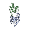
| ||||||||
|---|---|---|---|---|---|---|---|---|---|
| 1 | 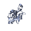
| ||||||||
| 2 | 
| ||||||||
| Unit cell |
|
-Components
| #1: Protein | Mass: 14872.784 Da / Num. of mol.: 2 / Fragment: P60 DOMAIN Source method: isolated from a genetically manipulated source Source: (gene. exp.) Mycobacterium avium subsp. paratuberculosis (bacteria)Strain: K-10 / Gene: MAP1204, MAP_1204 / Plasmid: pT7HMT / Production host: References: UniProt: Q740Y8, gamma-D-glutamyl-meso-diaminopimelate peptidase #2: Chemical | ChemComp-IPA / |   Mass: 60.095 Da / Num. of mol.: 1 / Source method: obtained synthetically / Formula: C3H8O / Comment: alkaloid*YM Mass: 60.095 Da / Num. of mol.: 1 / Source method: obtained synthetically / Formula: C3H8O / Comment: alkaloid*YM#3: Water | ChemComp-HOH / |  Mass: 18.015 Da / Num. of mol.: 45 / Source method: isolated from a natural source / Formula: H2O Mass: 18.015 Da / Num. of mol.: 45 / Source method: isolated from a natural source / Formula: H2O |
|---|
-Experimental details
-Experiment
| Experiment | Method: X-RAY DIFFRACTION / Number of used crystals: 1 |
|---|
- Sample preparation
| Crystal | Density Matthews: 2.57 Å3/Da / Density % sol: 52.05 % |
|---|---|
| Crystal grow | Temperature: 277 K / Method: vapor diffusion, hanging drop / pH: 7.5 Details: 22% PEG 3350, 18% 2-propanol, 0.1M NaHEPES, pH 7.5, VAPOR DIFFUSION, HANGING DROP, temperature 277K |
-Data collection
| Diffraction | Mean temperature: 100 K |
|---|---|
| Diffraction source | Source: SYNCHROTRON / Site: APS  / Beamline: 22-BM / Wavelength: 1 Å / Beamline: 22-BM / Wavelength: 1 Å |
| Detector | Type: MARMOSAIC 225 mm CCD / Detector: CCD / Date: Feb 12, 2009 |
| Radiation | Protocol: SINGLE WAVELENGTH / Monochromatic (M) / Laue (L): M / Scattering type: x-ray |
| Radiation wavelength | Wavelength: 1 Å / Relative weight: 1 |
| Reflection | Resolution: 2.4→43.43 Å / Num. obs: 12254 / % possible obs: 99.6 % / Observed criterion σ(F): 2.8 / Observed criterion σ(I): 5.6 / Redundancy: 13 % / Biso Wilson estimate: 39.333 Å2 / Rmerge(I) obs: 0.169 / Rsym value: 0.199 / Net I/σ(I): 14.09 |
| Reflection shell | Resolution: 2.4→2.49 Å / Redundancy: 7.4 % / Rmerge(I) obs: 0.856 / Mean I/σ(I) obs: 2.2 / Num. unique all: 1213 / Rsym value: 0.856 / % possible all: 100 |
- Processing
| Software |
| |||||||||||||||||||||||||||||||||||
|---|---|---|---|---|---|---|---|---|---|---|---|---|---|---|---|---|---|---|---|---|---|---|---|---|---|---|---|---|---|---|---|---|---|---|---|---|
| Refinement | Method to determine structure: MOLECULAR REPLACEMENT Starting model: PDB ENTRY 3GT2 Resolution: 2.4→43.428 Å / SU ML: 1.11 / Isotropic thermal model: ISOTROPIC / Cross valid method: THROUGHT / σ(F): 1.34 / σ(I): 3.9 / Stereochemistry target values: ML / Details: USED PHENIX.REFINE
| |||||||||||||||||||||||||||||||||||
| Solvent computation | Shrinkage radii: 0.9 Å / VDW probe radii: 1.11 Å / Solvent model: FLAT BULK SOLVENT MODEL / Bsol: 30.938 Å2 / ksol: 0.297 e/Å3 | |||||||||||||||||||||||||||||||||||
| Displacement parameters | Biso mean: 30.21 Å2
| |||||||||||||||||||||||||||||||||||
| Refinement step | Cycle: LAST / Resolution: 2.4→43.428 Å
| |||||||||||||||||||||||||||||||||||
| Refine LS restraints |
| |||||||||||||||||||||||||||||||||||
| LS refinement shell |
|
