 Movie
Movie Controller

[English] 日本語
 Yorodumi
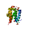
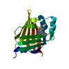
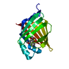
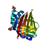
- PDB-4p15: Structure of the ClpC N-terminal domain from an alkaliphilic Baci... -
+ Open data

- Basic information
| Entry | Database: PDB / ID: 4p15 | ||||||
|---|---|---|---|---|---|---|---|
| Title | Structure of the ClpC N-terminal domain from an alkaliphilic Bacillus lehensis G1 species | ||||||
 Components Components | Bacillus lehensis ClpC N-terminal domain | ||||||
 Keywords Keywords | HYDROLASE / Heat-shock protein / ClpC / Bacillus / alkalophilic | ||||||
| Function / homology |  Function and homology information Function and homology informationATP-dependent Clp protease ATP-binding subunit CLPT1/2 / Double Clp-N motif / Clp, N-terminal domain / Clp repeat (R) N-terminal domain / Clp repeat (R) domain profile. / Clp, repeat (R) domain / Clp, N-terminal domain superfamily / Orthogonal Bundle / Mainly Alpha Similarity search - Domain/homology | ||||||
| Biological species |  Bacillus lehensis (bacteria) Bacillus lehensis (bacteria) | ||||||
| Method |  X-RAY DIFFRACTION / MOLECULAR REPLACEMENT / Resolution: 1.85 Å X-RAY DIFFRACTION / MOLECULAR REPLACEMENT / Resolution: 1.85 Å | ||||||
 Authors Authors | Rashid, S.A. / Littler, D.R. / Illias, R.M. / Murad, A.M.A. / Rossjohn, J. / Beddoe, T. / Mahadi, N.M. | ||||||
| Funding support |  Malaysia, 1items Malaysia, 1items
| ||||||
 Citation Citation | Journal: To Be Published Title: Structure of the ClpC N-terminal domain at 1.85 Angstroms resolution from an alkaliphilic Bacillus lehensis G1 species Authors: Rashid, S.A. / Littler, D.R. | ||||||
| History |
|
- Structure visualization
| Structure viewer | Molecule: MolmilJmol/JSmol |
|---|
- Downloads & links
-Download
| PDBx/mmCIF format | 4p15.cif.gz | 39.1 KB | Display | PDBx/mmCIF format |
|---|---|---|---|---|
| PDB format | pdb4p15.ent.gz | 26.3 KB | Display | PDB format |
| PDBx/mmJSON format | 4p15.json.gz | Tree view | PDBx/mmJSON format | |
| Others |  Other downloads Other downloads |
-Validation report
| Arichive directory | https://data.pdbj.org/pub/pdb/validation_reports/p1/4p15ftp://data.pdbj.org/pub/pdb/validation_reports/p1/4p15 | HTTPS FTP |
|---|
-Related structure data
| Similar structure data |
|---|


-Links
 PDBj
PDBj- Assembly
| Deposited unit | 
| ||||||||
|---|---|---|---|---|---|---|---|---|---|
| 1 |
| ||||||||
| Unit cell |
|
-Components
| #1: Protein | Mass: 15891.183 Da / Num. of mol.: 1 Source method: isolated from a genetically manipulated source Source: (gene. exp.) Bacillus lehensis (bacteria) / Production host: | ||
|---|---|---|---|
| #2: Chemical |   Mass: 96.063 Da / Num. of mol.: 2 / Source method: obtained synthetically / Formula: SO4 Mass: 96.063 Da / Num. of mol.: 2 / Source method: obtained synthetically / Formula: SO4#3: Water | ChemComp-HOH / |  Mass: 18.015 Da / Num. of mol.: 12 / Source method: isolated from a natural source / Formula: H2O Mass: 18.015 Da / Num. of mol.: 12 / Source method: isolated from a natural source / Formula: H2O |
-Experimental details
-Experiment
| Experiment | Method: X-RAY DIFFRACTION / Number of used crystals: 1 |
|---|
- Sample preparation
| Crystal | Density Matthews: 2.22 Å3/Da / Density % sol: 44.49 % / Description: Hexagonal crystal |
|---|---|
| Crystal grow | Temperature: 292.15 K / Method: vapor diffusion, hanging drop / pH: 4.2 Details: Protein crystallization was carried out using the hanging-drop vapor-diffusion method using 24-well Limbro tissue culture plates (ICN Inc.) at 19C. Drops were formed by mixing equal volumes ...Details: Protein crystallization was carried out using the hanging-drop vapor-diffusion method using 24-well Limbro tissue culture plates (ICN Inc.) at 19C. Drops were formed by mixing equal volumes (1 ul) of protein solution at 50 mg/mL and the reservoir solution containing 0.1M phosphate-citrate pH 4.2 20% polyethylene glycol 1000, 0.2M lithium sulphate. Hexagonal crystals appeared after 20-30 days |
-Data collection
| Diffraction | Mean temperature: 100 K |
|---|---|
| Diffraction source | Source: ROTATING ANODE / Type: RIGAKU MICROMAX-007 HF / Wavelength: 1.54178 Å |
| Detector | Type: RIGAKU RAXIS IV++ / Detector: IMAGE PLATE / Date: Dec 6, 2012 / Details: Osmic focusing mirrors |
| Radiation | Protocol: SINGLE WAVELENGTH / Monochromatic (M) / Laue (L): M / Scattering type: x-ray |
| Radiation wavelength | Wavelength: 1.54178 Å / Relative weight: 1 |
| Reflection | Resolution: 1.85→73.29 Å / Num. all: 11473 / Num. obs: 11473 / % possible obs: 99.6 % / Redundancy: 6.85 % / Rmerge(I) obs: 0.058 / Net I/σ(I): 19.6 |
| Reflection shell | Resolution: 1.85→73.29 Å / Redundancy: 6.85 % / Rmerge(I) obs: 0.058 / Mean I/σ(I) obs: 19.6 / % possible all: 99.6 |
- Processing
| Software | Name: PHENIX / Version: (phenix.refine: 1.8.2_1309) / Classification: refinement | |||||||||||||||||||||||||||||||||||
|---|---|---|---|---|---|---|---|---|---|---|---|---|---|---|---|---|---|---|---|---|---|---|---|---|---|---|---|---|---|---|---|---|---|---|---|---|
| Refinement | Method to determine structure: MOLECULAR REPLACEMENT / Resolution: 1.85→36.641 Å / SU ML: 0.21 / Cross valid method: THROUGHOUT / σ(F): 1.34 / Phase error: 26.82 / Stereochemistry target values: ML
| |||||||||||||||||||||||||||||||||||
| Solvent computation | Shrinkage radii: 0.9 Å / VDW probe radii: 1.11 Å / Solvent model: FLAT BULK SOLVENT MODEL | |||||||||||||||||||||||||||||||||||
| Refinement step | Cycle: 1 / Resolution: 1.85→36.641 Å
| |||||||||||||||||||||||||||||||||||
| Refine LS restraints |
| |||||||||||||||||||||||||||||||||||
| LS refinement shell |
|
