protein localization to perinuclear region of cytoplasm / brown fat cell proliferation / protein targeting to vacuole involved in autophagy / regulation of Ras protein signal transduction / intracellular membraneless organelle / aggrephagy / response to mitochondrial depolarisation / negative regulation of toll-like receptor 4 signaling pathway / amphisome / regulation of protein complex stability ...protein localization to perinuclear region of cytoplasm / brown fat cell proliferation / protein targeting to vacuole involved in autophagy / regulation of Ras protein signal transduction / intracellular membraneless organelle / aggrephagy / response to mitochondrial depolarisation / negative regulation of toll-like receptor 4 signaling pathway / amphisome / regulation of protein complex stability / endosome organization / autophagy of mitochondrion / pexophagy / membraneless organelle assembly / phagophore assembly site / regulation of mitochondrion organization / ubiquitin-modified protein reader activity / regulation of canonical NF-kappaB signal transduction / Nuclear events mediated by NFE2L2 / aggresome / endosomal transport / K63-linked polyubiquitin modification-dependent protein binding / cellular response to stress / Lewy body / temperature homeostasis / autolysosome / negative regulation of ferroptosis / molecular sequestering activity / immune system process / mitophagy / energy homeostasis / signaling adaptor activity / inclusion body / negative regulation of protein ubiquitination / positive regulation of autophagy / autophagosome / SH2 domain binding / ionotropic glutamate receptor binding / p75NTR recruits signalling complexes / NF-kB is activated and signals survival / Pexophagy / NRIF signals cell death from the nucleus / response to ischemia / protein kinase C binding / positive regulation of long-term synaptic potentiation / protein catabolic process / PINK1-PRKN Mediated Mitophagy / sarcomere / positive regulation of protein localization to plasma membrane / macroautophagy / ubiquitin binding / P-body / protein sequestering activity / molecular condensate scaffold activity / receptor tyrosine kinase binding / PML body / autophagy / Interleukin-1 signaling / protein import into nucleus / Signaling by ALK fusions and activated point mutants / intracellular protein localization / late endosome / KEAP1-NFE2L2 pathway / Neddylation / signaling receptor activity / sperm midpiece / transcription by RNA polymerase II / ubiquitin-dependent protein catabolic process / protein-macromolecule adaptor activity / cell differentiation / intracellular signal transduction / positive regulation of apoptotic process / apoptotic process / ubiquitin protein ligase binding / protein kinase binding / protein-containing complex binding / glutamatergic synapse / enzyme binding / negative regulation of transcription by RNA polymerase II / endoplasmic reticulum / positive regulation of transcription by RNA polymerase II / mitochondrion / extracellular exosome / zinc ion binding / nucleoplasm / identical protein binding / cytoplasm / cytosol Similarity search - Function
Sequestosome-1 / Phosphotyrosine-independent ligand for the Lck SH2 domain of 62 kDa / Ubiquitin-binding protein p62 ...Phosphotyrosine-independent ligand for the Lck SH2 domain of 62 kDa / Ubiquitin-binding protein p62 / EBI3-associated protein of 60 kDa / p60 / EBIAP
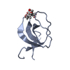
Mass: 5744.406 Da / Num. of mol.: 1 / Fragment: UBA domain (UNP residues 387-436) Source method: isolated from a genetically manipulated source Source: (gene. exp.) Homo sapiens (human) / Gene: SQSTM1, ORCA, OSIL / Plasmid: pGEX-4T-1 / Species (production host): Escherichia coli / Production host: Escherichia coli BL21(DE3) (bacteria) / Strain (production host): BL21(DE3) / References: UniProt: Q13501
-
Experimental details
-
Experiment
Experiment
Method: SOLUTION NMR
NMR experiment
Conditions-ID
Experiment-ID
Solution-ID
Type
1
1
1
2D 1H-15N HSQC
1
2
1
3D 1H-15N NOESY
1
3
2
2D 1H-13C HSQC
1
4
2
3DCBCA(CO)NH
1
5
2
3D HN(CA)CB
1
6
2
3D HNCO
1
7
2
3DHN(CA)CO
1
8
2
3D (H)CCH-TOCSY
1
9
2
3D 1H-13C NOESY
1
10
1
3D 1H-15N TOCSY
-
Sample preparation
Details
Solution-ID
Contents
Solvent system
1
1 mM [U-100% 15N] Sequestosome-1, 50 mM potassium phosphate, 50 mM sodium chloride, 90% H2O/10% D2O
90% H2O/10% D2O
2
1 mM [U-100% 13C; U-100% 15N] Sequestosome-1, 50 mM potassium phosphate, 50 mM sodium chloride, 90% H2O/10% D2O
90% H2O/10% D2O
Sample
Conc. (mg/ml)
Component
Isotopic labeling
Solution-ID
1mM
Sequestosome-1
[U-100% 15N]
1
50mM
potassiumphosphate
1
50mM
sodiumchloride
1
1mM
Sequestosome-1
[U-100% 13C; U-100% 15N]
2
50mM
potassiumphosphate
2
50mM
sodiumchloride
2
Sample conditions
pH: 7 / Pressure: ambient atm / Temperature: 298 K
-
NMR measurement
NMR spectrometer
Type: Bruker Avance / Manufacturer: Bruker / Model: AVANCE / Field strength: 600 MHz
-
Processing
NMR software
Name
Version
Developer
Classification
X-PLOR NIH
2.14
Schwieters, Kuszewski, TjandraandClore
structuresolution
TALOS
Cornilescu, DelaglioandBax
dataanalysis
XwinNMR
BrukerBiospin
collection
XwinNMR
BrukerBiospin
processing
CcpNMR
1.0.10
CCPN
chemicalshiftassignment
CcpNMR
1.0.10
CCPN
dataanalysis
CcpNMR
1.0.10
CCPN
peakpicking
X-PLOR NIH
2.14
Schwieters, Kuszewski, TjandraandClore
refinement
Refinement
Method: simulated annealing / Software ordinal: 1
NMR constraints
NOE constraints total: 1078 / NOE intraresidue total count: 360 / NOE long range total count: 213 / NOE medium range total count: 246 / NOE sequential total count: 259 / Protein phi angle constraints total count: 36 / Protein psi angle constraints total count: 36
NMR representative
Selection criteria: closest to the average
NMR ensemble
Conformer selection criteria: structures with the lowest energy Conformers calculated total number: 100 / Conformers submitted total number: 30
+
About Yorodumi
-
News
-
Feb 9, 2022. New format data for meta-information of EMDB entries
New format data for meta-information of EMDB entries
Version 3 of the EMDB header file is now the official format.
The previous official version 1.9 will be removed from the archive.
In the structure databanks used in Yorodumi, some data are registered as the other names, "COVID-19 virus" and "2019-nCoV". Here are the details of the virus and the list of structure data.
Jan 31, 2019. EMDB accession codes are about to change! (news from PDBe EMDB page)
EMDB accession codes are about to change! (news from PDBe EMDB page)
The allocation of 4 digits for EMDB accession codes will soon come to an end. Whilst these codes will remain in use, new EMDB accession codes will include an additional digit and will expand incrementally as the available range of codes is exhausted. The current 4-digit format prefixed with “EMD-” (i.e. EMD-XXXX) will advance to a 5-digit format (i.e. EMD-XXXXX), and so on. It is currently estimated that the 4-digit codes will be depleted around Spring 2019, at which point the 5-digit format will come into force.
The EM Navigator/Yorodumi systems omit the EMD- prefix.
Related info.:Q: What is EMD? / ID/Accession-code notation in Yorodumi/EM Navigator
Yorodumi is a browser for structure data from EMDB, PDB, SASBDB, etc.
This page is also the successor to EM Navigator detail page, and also detail information page/front-end page for Omokage search.
The word "yorodu" (or yorozu) is an old Japanese word meaning "ten thousand". "mi" (miru) is to see.
Related info.:EMDB / PDB / SASBDB / Comparison of 3 databanks / Yorodumi Search / Aug 31, 2016. New EM Navigator & Yorodumi / Yorodumi Papers / Jmol/JSmol / Function and homology information / Changes in new EM Navigator and Yorodumi
 Movie
Movie Controller


 Open data
 Basic information
 Components
 Keywords
 Function and homology information
 Homo sapiens (human)
 Authors
 Citation
 Structure visualization
 Downloads & links
 Other downloads
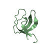


 PDBj
PDBj


 Assembly
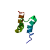

 HSQC
HSQC Sample preparation
 Processing