 Movie
Movie Controller

[English] 日本語
 Yorodumi
- PDB-2jg1: STRUCTURE OF Staphylococcus aureus D-TAGATOSE-6-PHOSPHATE KINASE ... -
+ Open data

- Basic information
| Entry | Database: PDB / ID: 2jg1 | ||||||
|---|---|---|---|---|---|---|---|
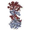
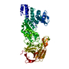
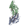
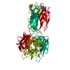
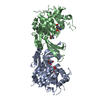
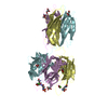
| Title | STRUCTURE OF Staphylococcus aureus D-TAGATOSE-6-PHOSPHATE KINASE with cofactor and substrate | ||||||
 Components Components | TAGATOSE-6-PHOSPHATE KINASE | ||||||
 Keywords Keywords | TRANSFERASE / D-TAGATOSE-6-PHOSPHATE KINASE / PHOSPHORYL TRANSFER / CONFORMATIONAL CHANGES / KINASE / LACTOSE METABOLISM | ||||||
| Function / homology |  Function and homology information Function and homology informationtagatose-6-phosphate kinase / : / phosphofructokinase activity / D-tagatose 6-phosphate catabolic process / tagatose-6-phosphate kinase activity / small molecule metabolic process / ATP binding / cytosol Similarity search - Function | ||||||
| Biological species |   STAPHYLOCOCCUS AUREUS (bacteria) STAPHYLOCOCCUS AUREUS (bacteria) | ||||||
| Method |  X-RAY DIFFRACTION / SYNCHROTRON / SAD / Resolution: 2 Å X-RAY DIFFRACTION / SYNCHROTRON / SAD / Resolution: 2 Å | ||||||
 Authors Authors | Miallau, L. / Hunter, W.N. / McSweeney, S.M. / Leonard, G.A. | ||||||
 Citation Citation | Journal: J.Biol.Chem. / Year: 2007 Title: Structures of Staphylococcus Aureus D-Tagatose-6-Phosphate Kinase Implicate Domain Motions in Specificity and Mechanism. Authors: Miallau, L. / Hunter, W.N. / Mcsweeney, S.M. / Leonard, G.A. | ||||||
| History |
| ||||||
| Remark 700 | SHEET THE SHEET STRUCTURE OF THIS MOLECULE IS BIFURCATED. IN ORDER TO REPRESENT THIS FEATURE IN ... SHEET THE SHEET STRUCTURE OF THIS MOLECULE IS BIFURCATED. IN ORDER TO REPRESENT THIS FEATURE IN THE SHEET RECORDS BELOW, TWO SHEETS ARE DEFINED. |
- Structure visualization
| Structure viewer | Molecule: MolmilJmol/JSmol |
|---|
- Downloads & links
-Download
| PDBx/mmCIF format | 2jg1.cif.gz | 276.9 KB | Display | PDBx/mmCIF format |
|---|---|---|---|---|
| PDB format | pdb2jg1.ent.gz | 224.5 KB | Display | PDB format |
| PDBx/mmJSON format | 2jg1.json.gz | Tree view | PDBx/mmJSON format | |
| Others |  Other downloads Other downloads |
-Validation report
| Arichive directory | https://data.pdbj.org/pub/pdb/validation_reports/jg/2jg1ftp://data.pdbj.org/pub/pdb/validation_reports/jg/2jg1 | HTTPS FTP |
|---|
-Related structure data

-Links
 PDBj
PDBj


- Assembly
| Deposited unit | 
| ||||||||||||
|---|---|---|---|---|---|---|---|---|---|---|---|---|---|
| 1 | 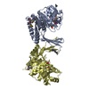
| ||||||||||||
| 2 | 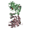
| ||||||||||||
| Unit cell |
| ||||||||||||
| Noncrystallographic symmetry (NCS) | NCS oper:
|
-Components
| #1: Protein | Mass: 36325.184 Da / Num. of mol.: 4 / Mutation: YES Source method: isolated from a genetically manipulated source Source: (gene. exp.) STAPHYLOCOCCUS AUREUS (bacteria) / Strain: NCTC 8325 / Production host: #2: Chemical | ChemComp-MG /   Mass: 24.305 Da / Num. of mol.: 5 / Source method: obtained synthetically / Formula: Mg Mass: 24.305 Da / Num. of mol.: 5 / Source method: obtained synthetically / Formula: Mg#3: Chemical | ChemComp-ANP /   Mass: 506.196 Da / Num. of mol.: 4 / Source method: obtained synthetically / Formula: C10H17N6O12P3 / Comment: AMP-PNP, energy-carrying molecule analogue*YM Mass: 506.196 Da / Num. of mol.: 4 / Source method: obtained synthetically / Formula: C10H17N6O12P3 / Comment: AMP-PNP, energy-carrying molecule analogue*YM#4: Sugar | ChemComp-TA6 / |   Type: D-saccharide, beta linking / Mass: 260.136 Da / Num. of mol.: 1 / Source method: obtained synthetically / Formula: C6H13O9P Type: D-saccharide, beta linking / Mass: 260.136 Da / Num. of mol.: 1 / Source method: obtained synthetically / Formula: C6H13O9P#5: Water | ChemComp-HOH / |  Mass: 18.015 Da / Num. of mol.: 875 / Source method: isolated from a natural source / Formula: H2O Mass: 18.015 Da / Num. of mol.: 875 / Source method: isolated from a natural source / Formula: H2OCompound details | ENGINEERED RESIDUE IN CHAIN A, LEU 124 TO MSE ENGINEERED RESIDUE IN CHAIN A, LEU 125 TO MSE ...ENGINEERED | Has protein modification | Y | |
|---|
-Experimental details
-Experiment
| Experiment | Method: X-RAY DIFFRACTION / Number of used crystals: 1 |
|---|
- Sample preparation
| Crystal | Density Matthews: 2.3 Å3/Da / Density % sol: 50 % |
|---|---|
| Crystal grow | pH: 6.5 Details: 0.2 M MAGNESIUM ACETATE TETRAHYDRATE, 0.1 M SODIUM CACODYLATE PH 6.5, 10% PEG 8000, 15% PEG 550, 10 MM ATP. |
-Data collection
| Diffraction | Mean temperature: 100 K |
|---|---|
| Diffraction source | Source: SYNCHROTRON / Site: ESRF  / Beamline: ID14-2 / Wavelength: 0.933 / Beamline: ID14-2 / Wavelength: 0.933 |
| Detector | Type: ADSC CCD / Detector: CCD |
| Radiation | Protocol: SINGLE WAVELENGTH / Monochromatic (M) / Laue (L): M / Scattering type: x-ray |
| Radiation wavelength | Wavelength: 0.933 Å / Relative weight: 1 |
| Reflection | Resolution: 2→24.8 Å / Num. obs: 96948 / % possible obs: 99.8 % / Redundancy: 10.9 % / Rmerge(I) obs: 0.09 / Net I/σ(I): 7.6 |
| Reflection shell | Resolution: 2→2.12 Å / Redundancy: 9.6 % / Rmerge(I) obs: 0.62 / Mean I/σ(I) obs: 1.2 / % possible all: 99.8 |
- Processing
| Software |
| ||||||||||||||||||||||||||||||||||||||||||||||||||||||||||||||||||||||||||||||||||||||||||||||||||||||||||||||||||||||||||||||||||||||||||||||||||||||||||||||||||||||||||||||||||||||
|---|---|---|---|---|---|---|---|---|---|---|---|---|---|---|---|---|---|---|---|---|---|---|---|---|---|---|---|---|---|---|---|---|---|---|---|---|---|---|---|---|---|---|---|---|---|---|---|---|---|---|---|---|---|---|---|---|---|---|---|---|---|---|---|---|---|---|---|---|---|---|---|---|---|---|---|---|---|---|---|---|---|---|---|---|---|---|---|---|---|---|---|---|---|---|---|---|---|---|---|---|---|---|---|---|---|---|---|---|---|---|---|---|---|---|---|---|---|---|---|---|---|---|---|---|---|---|---|---|---|---|---|---|---|---|---|---|---|---|---|---|---|---|---|---|---|---|---|---|---|---|---|---|---|---|---|---|---|---|---|---|---|---|---|---|---|---|---|---|---|---|---|---|---|---|---|---|---|---|---|---|---|---|---|
| Refinement | Method to determine structure: SAD / Resolution: 2→24.85 Å / Cor.coef. Fo:Fc: 0.953 / Cor.coef. Fo:Fc free: 0.935 / SU B: 4.131 / SU ML: 0.117 / Cross valid method: THROUGHOUT / ESU R: 0.183 / ESU R Free: 0.164 / Stereochemistry target values: MAXIMUM LIKELIHOOD / Details: HYDROGENS HAVE BEEN ADDED IN THE RIDING POSITIONS
| ||||||||||||||||||||||||||||||||||||||||||||||||||||||||||||||||||||||||||||||||||||||||||||||||||||||||||||||||||||||||||||||||||||||||||||||||||||||||||||||||||||||||||||||||||||||
| Solvent computation | Ion probe radii: 0.8 Å / Shrinkage radii: 0.8 Å / VDW probe radii: 1.4 Å / Solvent model: MASK | ||||||||||||||||||||||||||||||||||||||||||||||||||||||||||||||||||||||||||||||||||||||||||||||||||||||||||||||||||||||||||||||||||||||||||||||||||||||||||||||||||||||||||||||||||||||
| Displacement parameters | Biso mean: 31.09 Å2
| ||||||||||||||||||||||||||||||||||||||||||||||||||||||||||||||||||||||||||||||||||||||||||||||||||||||||||||||||||||||||||||||||||||||||||||||||||||||||||||||||||||||||||||||||||||||
| Refinement step | Cycle: LAST / Resolution: 2→24.85 Å
| ||||||||||||||||||||||||||||||||||||||||||||||||||||||||||||||||||||||||||||||||||||||||||||||||||||||||||||||||||||||||||||||||||||||||||||||||||||||||||||||||||||||||||||||||||||||
| Refine LS restraints |
|