Mass: 18.015 Da / Num. of mol.: 67 / Source method: isolated from a natural source / Formula: H2O
Sequence details
GSH AT START OF SEQUENCE IS A REMNANT OF THE N-TERMINAL THROMBIN CLEAVED 6XHIS-TAG USED IN PURIFICATION.
-
Experimental details
-
Experiment
Experiment
Method: X-RAY DIFFRACTION / Number of used crystals: 1
-
Sample preparation
Crystal
Density Matthews: 2.61 Å3/Da / Density % sol: 52.9 %
Crystal grow
Method: vapor diffusion, hanging drop / pH: 7.5 Details: PROTEIN AT 15MG/ML IN 20 MM TRIS PH 8.0 CONTAINING 1 MM DTT, 0.1 MM EDTA, 4MM N1-ACETYLSPERMINE-S-COA. CRYSTALLIZED BY HANGING DROP VAPOUR DIFFUSION WITH 3-4 M NACL, 200 MM K/NA TARTRATE, ...Details: PROTEIN AT 15MG/ML IN 20 MM TRIS PH 8.0 CONTAINING 1 MM DTT, 0.1 MM EDTA, 4MM N1-ACETYLSPERMINE-S-COA. CRYSTALLIZED BY HANGING DROP VAPOUR DIFFUSION WITH 3-4 M NACL, 200 MM K/NA TARTRATE, 100 MM TRIS-HCL PH 7-8, 50 MM MGCL2
Resolution: 2.3→47.95 Å / Cor.coef. Fo:Fc: 0.941 / Cor.coef. Fo:Fc free: 0.915 / SU B: 8.647 / SU ML: 0.214 / Cross valid method: THROUGHOUT / ESU R: 0.405 / ESU R Free: 0.271 / Stereochemistry target values: MAXIMUM LIKELIHOOD / Details: HYDROGENS HAVE BEEN ADDED IN THE RIDING POSITIONS.
Rfactor
Num. reflection
% reflection
Selection details
Rfree
0.275
908
5.1 %
RANDOM
Rwork
0.224
-
-
-
obs
0.227
16978
98.3 %
-
Solvent computation
Ion probe radii: 0.8 Å / Shrinkage radii: 0.8 Å / VDW probe radii: 1.2 Å / Solvent model: MASK
 Movie
Movie Controller

 Yorodumi
 Open data
 Basic information
 Components
 Keywords
 Function and homology information
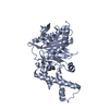
 HOMO SAPIENS (human)
 X-RAY DIFFRACTION /
  Authors
 Citation
 Structure visualization
 Downloads & links
 Other downloads


 PDBj
PDBj

 Assembly

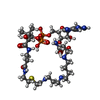
 Mass: 1009.895 Da / Num. of mol.: 2 / Source method: obtained synthetically / Formula: C33H62N11O17P3S
 Mass: 18.015 Da / Num. of mol.: 67 / Source method: isolated from a natural source / Formula: H2O
 Sample preparation
 Processing