 Movie
Movie Controller


+ Open data

- Basic information
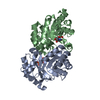
| Entry | Database: PDB / ID: 2b5g | ||||||
|---|---|---|---|---|---|---|---|
| Title | Wild Type SSAT- 1.7A structure | ||||||
 Components Components | (Diamine acetyltransferase 1) x 2 | ||||||
 Keywords Keywords | TRANSFERASE / Structural Genomics / PSI / Protein Structure Initiative / New York SGX Research Center for Structural Genomics / NYSGXRC | ||||||
| Function / homology |  Function and homology information Function and homology informationInterconversion of polyamines / : / putrescine catabolic process / spermidine binding / polyamine biosynthetic process / diamine N-acetyltransferase / diamine N-acetyltransferase activity / N-acetyltransferase activity / regulation of cell population proliferation / angiogenesis ...Interconversion of polyamines / : / putrescine catabolic process / spermidine binding / polyamine biosynthetic process / diamine N-acetyltransferase / diamine N-acetyltransferase activity / N-acetyltransferase activity / regulation of cell population proliferation / angiogenesis / identical protein binding / cytosol Similarity search - Function | ||||||
| Biological species |  Homo sapiens (human) Homo sapiens (human) | ||||||
| Method |  X-RAY DIFFRACTION / SAD / Resolution: 1.7 Å X-RAY DIFFRACTION / SAD / Resolution: 1.7 Å | ||||||
 Authors Authors | Bewley, M.C. / Graziano, V. / Jiang, J.S. / Matz, E. / Studier, F.W. / Pegg, A.P. / Coleman, C.S. / Flanagan, J.M. / Burley, S.K. / New York SGX Research Center for Structural Genomics (NYSGXRC) | ||||||
 Citation Citation | Journal: Proc.Natl.Acad.Sci.Usa / Year: 2006 Title: Structures of wild-type and mutant human spermidine/spermine N1-acetyltransferase, a potential therapeutic drug target Authors: Bewley, M.C. / Graziano, V. / Jiang, J.S. / Matz, E. / Studier, F.W. / Pegg, A.P. / Coleman, C.S. / Flanagan, J.M. | ||||||
| History |
|
- Structure visualization
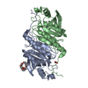
| Structure viewer | Molecule: MolmilJmol/JSmol |
|---|
- Downloads & links
-Download
| PDBx/mmCIF format | 2b5g.cif.gz | 86.7 KB | Display | PDBx/mmCIF format |
|---|---|---|---|---|
| PDB format | pdb2b5g.ent.gz | 65.8 KB | Display | PDB format |
| PDBx/mmJSON format | 2b5g.json.gz | Tree view | PDBx/mmJSON format | |
| Others |  Other downloads Other downloads |
-Validation report
| Arichive directory | https://data.pdbj.org/pub/pdb/validation_reports/b5/2b5gftp://data.pdbj.org/pub/pdb/validation_reports/b5/2b5g | HTTPS FTP |
|---|
-Related structure data
| Related structure data |  2b3uC  2b3vC  2b4bC  2b4dC  2b58C C: citing same article ( |
|---|---|
| Similar structure data | |
| Other databases |


-Links
 PDBj
PDBj

- Assembly
| Deposited unit | 
| ||||||||
|---|---|---|---|---|---|---|---|---|---|
| 1 |
| ||||||||
| Unit cell |
|
-Components
| #1: Protein | Mass: 20376.254 Da / Num. of mol.: 1 Source method: isolated from a genetically manipulated source Source: (gene. exp.) Homo sapiens (human) / Gene: SAT / Production host:  | ||||
|---|---|---|---|---|---|
| #2: Protein | Mass: 20417.283 Da / Num. of mol.: 1 Source method: isolated from a genetically manipulated source References: UniProt: P21673 | ||||
| #3: Chemical | ChemComp-SO4 /   Mass: 96.063 Da / Num. of mol.: 7 / Source method: obtained synthetically / Formula: SO4 Mass: 96.063 Da / Num. of mol.: 7 / Source method: obtained synthetically / Formula: SO4#4: Water | ChemComp-HOH / |  Mass: 18.015 Da / Num. of mol.: 323 / Source method: isolated from a natural source / Formula: H2O Mass: 18.015 Da / Num. of mol.: 323 / Source method: isolated from a natural source / Formula: H2OHas protein modification | Y | |
-Experimental details
-Experiment
| Experiment | Method: X-RAY DIFFRACTION |
|---|
- Sample preparation
| Crystal | Density Matthews: 2.13 Å3/Da / Density % sol: 42.27 % |
|---|
-Data collection
| Radiation | Protocol: SINGLE WAVELENGTH / Monochromatic (M) / Laue (L): M / Scattering type: x-ray |
|---|---|
| Radiation wavelength | Relative weight: 1 |
- Processing
| Software | Name: CNS / Version: 1.1 / Classification: refinement | |||||||||||||||
|---|---|---|---|---|---|---|---|---|---|---|---|---|---|---|---|---|
| Refinement | Method to determine structure: SAD / Resolution: 1.7→30 Å / σ(F): -3
| |||||||||||||||
| Refinement step | Cycle: LAST / Resolution: 1.7→30 Å
|
