 Movie
Movie Controller

[English] 日本語
 Yorodumi
- PDB-2jb0: CRYSTAL STRUCTURE OF THE MUTANT H573A OF THE NUCLEASE DOMAIN OF C... -
+ Open data

- Basic information
| Entry | Database: PDB / ID: 2jb0 | ||||||
|---|---|---|---|---|---|---|---|
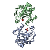
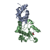
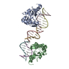
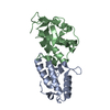
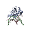
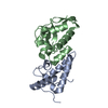
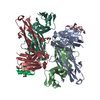
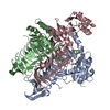
| Title | CRYSTAL STRUCTURE OF THE MUTANT H573A OF THE NUCLEASE DOMAIN OF COLE7 IN COMPLEX WITH IM7 | ||||||
 Components Components |
| ||||||
 Keywords Keywords | HYDROLASE/INHIBITOR / HYDROLASE-INHIBITOR COMPLEX / ZINC / TOXIN / PLASMID / NUCLEASE / HYDROLASE / ANTIBIOTIC / H-N-H MOTIF / BACTERIOCIN / ENDONUCLEASE / METAL-BINDING / ANTIMICROBIAL / DNA HYDROLYSIS / BACTERIOCIN IMMUNITY / HIS METAL FINGER MOTIF | ||||||
| Function / homology |  Function and homology information Function and homology informationextrachromosomal circular DNA / bacteriocin immunity / toxic substance binding / endonuclease activity / killing of cells of another organism / Hydrolases; Acting on ester bonds / defense response to bacterium / hydrolase activity / metal ion binding Similarity search - Function | ||||||
| Biological species |  | ||||||
| Method |  X-RAY DIFFRACTION / SYNCHROTRON / MOLECULAR REPLACEMENT / Resolution: 1.91 Å X-RAY DIFFRACTION / SYNCHROTRON / MOLECULAR REPLACEMENT / Resolution: 1.91 Å | ||||||
 Authors Authors | Huang, H. / Yuan, H.S. | ||||||
 Citation Citation | Journal: J.Mol.Biol. / Year: 2007 Title: The Conserved Asparagine in the Hnh Motif Serves an Important Structural Role in Metal Finger Endonucleases. Authors: Huang, H. / Yuan, H.S. | ||||||
| History |
|
- Structure visualization
| Structure viewer | Molecule: MolmilJmol/JSmol |
|---|
- Downloads & links
-Download
| PDBx/mmCIF format | 2jb0.cif.gz | 59.9 KB | Display | PDBx/mmCIF format |
|---|---|---|---|---|
| PDB format | pdb2jb0.ent.gz | 43.4 KB | Display | PDB format |
| PDBx/mmJSON format | 2jb0.json.gz | Tree view | PDBx/mmJSON format | |
| Others |  Other downloads Other downloads |
-Validation report
| Arichive directory | https://data.pdbj.org/pub/pdb/validation_reports/jb/2jb0ftp://data.pdbj.org/pub/pdb/validation_reports/jb/2jb0 | HTTPS FTP |
|---|
-Related structure data
| Related structure data | 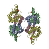 2jazC  2jbgC 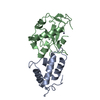 7ceiS S: Starting model for refinement C: citing same article ( |
|---|---|
| Similar structure data |


-Links
 PDBj
PDBj
- Assembly
| Deposited unit | 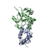
| ||||||||
|---|---|---|---|---|---|---|---|---|---|
| 1 | 
| ||||||||
| Unit cell |
|
-Components
| #1: Protein | Mass: 9906.963 Da / Num. of mol.: 1 Source method: isolated from a genetically manipulated source Source: (gene. exp.) References: UniProt: Q03708, Hydrolases; Acting on ester bonds |
|---|---|
| #2: Protein | Mass: 14995.036 Da / Num. of mol.: 1 / Fragment: NUCLEASE DOMAIN, RESIDUES 446-576 / Mutation: YES Source method: isolated from a genetically manipulated source Source: (gene. exp.) References: UniProt: Q47112, Hydrolases; Acting on ester bonds |
| #3: Chemical | ChemComp-ZN /   Mass: 65.409 Da / Num. of mol.: 1 / Source method: obtained synthetically / Formula: Zn Mass: 65.409 Da / Num. of mol.: 1 / Source method: obtained synthetically / Formula: Zn |
| #4: Water | ChemComp-HOH /  Mass: 18.015 Da / Num. of mol.: 193 / Source method: isolated from a natural source / Formula: H2O Mass: 18.015 Da / Num. of mol.: 193 / Source method: isolated from a natural source / Formula: H2O |
| Compound details | ENGINEERED |
-Experimental details
-Experiment
| Experiment | Method: X-RAY DIFFRACTION / Number of used crystals: 1 |
|---|
- Sample preparation
| Crystal | Density Matthews: 2.86 Å3/Da / Density % sol: 57.02 % |
|---|---|
| Crystal grow | pH: 7 Details: 20 % W/V PEG3350 AND 0.2 M DI-AMMONIUM HYDROGEN CITRATE, pH 7.00 |
-Data collection
| Diffraction | Mean temperature: 113 K |
|---|---|
| Diffraction source | Source: SYNCHROTRON / Site: SPring-8  / Beamline: BL12B2 / Wavelength: 1 / Beamline: BL12B2 / Wavelength: 1 |
| Detector | Type: ADSC CCD / Detector: CCD / Date: May 24, 2005 / Details: MIRRORS |
| Radiation | Monochromator: SI / Protocol: SINGLE WAVELENGTH / Monochromatic (M) / Laue (L): M / Scattering type: x-ray |
| Radiation wavelength | Wavelength: 1 Å / Relative weight: 1 |
| Reflection | Resolution: 1.91→25.46 Å / Num. obs: 21837 / % possible obs: 97.4 % / Observed criterion σ(I): 0 / Redundancy: 6.3 % / Biso Wilson estimate: 20.7 Å2 / Rsym value: 0.04 / Net I/σ(I): 39.2 |
| Reflection shell | Resolution: 1.91→1.98 Å / Redundancy: 3.5 % / Mean I/σ(I) obs: 4.8 / Rsym value: 0.27 / % possible all: 86.1 |
- Processing
| Software |
| ||||||||||||||||||||||||||||||||||||||||||||||||||||||||||||||||||||||||||||||||
|---|---|---|---|---|---|---|---|---|---|---|---|---|---|---|---|---|---|---|---|---|---|---|---|---|---|---|---|---|---|---|---|---|---|---|---|---|---|---|---|---|---|---|---|---|---|---|---|---|---|---|---|---|---|---|---|---|---|---|---|---|---|---|---|---|---|---|---|---|---|---|---|---|---|---|---|---|---|---|---|---|---|
| Refinement | Method to determine structure: MOLECULAR REPLACEMENT Starting model: PDB ENTRY 7CEI Resolution: 1.91→25.46 Å / Rfactor Rfree error: 0.005 / Data cutoff high absF: 506787.59 / Isotropic thermal model: RESTRAINED / Cross valid method: THROUGHOUT / σ(F): 0
| ||||||||||||||||||||||||||||||||||||||||||||||||||||||||||||||||||||||||||||||||
| Solvent computation | Solvent model: FLAT MODEL / Bsol: 53.7618 Å2 / ksol: 0.350635 e/Å3 | ||||||||||||||||||||||||||||||||||||||||||||||||||||||||||||||||||||||||||||||||
| Displacement parameters | Biso mean: 35.1 Å2
| ||||||||||||||||||||||||||||||||||||||||||||||||||||||||||||||||||||||||||||||||
| Refine analyze |
| ||||||||||||||||||||||||||||||||||||||||||||||||||||||||||||||||||||||||||||||||
| Refinement step | Cycle: LAST / Resolution: 1.91→25.46 Å
| ||||||||||||||||||||||||||||||||||||||||||||||||||||||||||||||||||||||||||||||||
| Refine LS restraints |
| ||||||||||||||||||||||||||||||||||||||||||||||||||||||||||||||||||||||||||||||||
| LS refinement shell | Resolution: 1.91→2.03 Å / Rfactor Rfree error: 0.016 / Total num. of bins used: 6
| ||||||||||||||||||||||||||||||||||||||||||||||||||||||||||||||||||||||||||||||||
| Xplor file |
|
