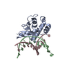
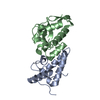
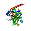
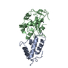
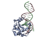
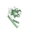
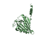
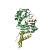
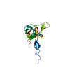
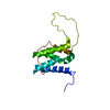
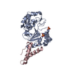
extrachromosomal circular DNA / endonuclease activity / killing of cells of another organism / Hydrolases; Acting on ester bonds / defense response to bacterium / hydrolase activity / metal ion binding Similarity search - Function
Mass: 18.015 Da / Num. of mol.: 87 / Source method: isolated from a natural source / Formula: H2O
Compound details
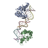
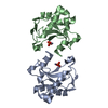
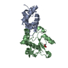
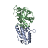
THIS PLASMID-CODED BACTERICIDAL PROTEIN IS AN ENDONUCLEASE ACTIVE ON BOTH SINGLE- AND DOUBLE- ...THIS PLASMID-CODED BACTERICIDAL PROTEIN IS AN ENDONUCLEASE ACTIVE ON BOTH SINGLE- AND DOUBLE-STRANDED DNA BUT WITH UNDEFINED SPECIFICITY. ENGINEERED RESIDUE IN CHAIN A, HIS 545 TO GLN
-
Experimental details
-
Experiment
Experiment
Method: X-RAY DIFFRACTION / Number of used crystals: 1
-
Sample preparation
Crystal
Density Matthews: 3.53 Å3/Da / Density % sol: 64.8 %
Crystal grow
Method: vapor diffusion, hanging drop / pH: 4.8 Details: HANGING DROP VAPOR DIFFUSION METHOD BY MIXING 1 MICRO L COMPLEX SOLUTION AND 1 MICRO L RESERVOIR SOLUTION CONSISTING OF 40 % MPD, 0.4 M AMMONIUM FORMATE AND 0.1 M ACETATE BUFFER (PH4.8) AT ...Details: HANGING DROP VAPOR DIFFUSION METHOD BY MIXING 1 MICRO L COMPLEX SOLUTION AND 1 MICRO L RESERVOIR SOLUTION CONSISTING OF 40 % MPD, 0.4 M AMMONIUM FORMATE AND 0.1 M ACETATE BUFFER (PH4.8) AT ROOM TEMPERATURE., pH 4.80
In the structure databanks used in Yorodumi, some data are registered as the other names, "COVID-19 virus" and "2019-nCoV". Here are the details of the virus and the list of structure data.
Jan 31, 2019. EMDB accession codes are about to change! (news from PDBe EMDB page)
EMDB accession codes are about to change! (news from PDBe EMDB page)
The allocation of 4 digits for EMDB accession codes will soon come to an end. Whilst these codes will remain in use, new EMDB accession codes will include an additional digit and will expand incrementally as the available range of codes is exhausted. The current 4-digit format prefixed with “EMD-” (i.e. EMD-XXXX) will advance to a 5-digit format (i.e. EMD-XXXXX), and so on. It is currently estimated that the 4-digit codes will be depleted around Spring 2019, at which point the 5-digit format will come into force.
The EM Navigator/Yorodumi systems omit the EMD- prefix.
Related info.:Q: What is EMD? / ID/Accession-code notation in Yorodumi/EM Navigator
Yorodumi is a browser for structure data from EMDB, PDB, SASBDB, etc.
This page is also the successor to EM Navigator detail page, and also detail information page/front-end page for Omokage search.
The word "yorodu" (or yorozu) is an old Japanese word meaning "ten thousand". "mi" (miru) is to see.
Related info.:EMDB / PDB / SASBDB / Comparison of 3 databanks / Yorodumi Search / Aug 31, 2016. New EM Navigator & Yorodumi / Yorodumi Papers / Jmol/JSmol / Function and homology information / Changes in new EM Navigator and Yorodumi
 Movie
Movie Controller

 Yorodumi
 Open data
 Basic information
 Components
 Keywords
 Function and homology information
 X-RAY DIFFRACTION /
  Authors
 Citation
 Structure visualization
 Downloads & links
 Other downloads


 PDBj
PDBj


 Assembly
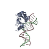

 Mass: 65.409 Da / Num. of mol.: 1 / Source method: obtained synthetically / Formula: Zn
 Mass: 18.015 Da / Num. of mol.: 87 / Source method: isolated from a natural source / Formula: H2O
 Sample preparation
 / Beamline: AR-NW12A / Wavelength: 1
  Processing