 Movie
Movie Controller

[English] 日本語
 Yorodumi
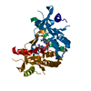
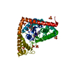
- PDB-2j8n: Structure of P. aeruginosa acetyltransferase PA4866 solved at roo... -
+ Open data

- Basic information
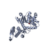
| Entry | Database: PDB / ID: 2j8n | ||||||
|---|---|---|---|---|---|---|---|
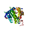
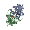
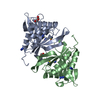
| Title | Structure of P. aeruginosa acetyltransferase PA4866 solved at room temperature | ||||||
 Components Components | ACETYLTRANSFERASE PA4866 FROM P. AERUGINOSA | ||||||
 Keywords Keywords | TRANSFERASE / GCN5 FAMILY / PHOSPHINOTHRICIN / METHIONINE SULFONE / PSEUDOMONAS AERUGINOSA / METHIONINE SULFOXIMINE / N-ACETYL TRANSFERASE / HYPOTHETICAL PROTEIN | ||||||
| Function / homology |  Function and homology information Function and homology informationacyltransferase activity, transferring groups other than amino-acyl groups / Transferases; Acyltransferases; Transferring groups other than aminoacyl groups Similarity search - Function | ||||||
| Biological species |   PSEUDOMONAS AERUGINOSA (bacteria) PSEUDOMONAS AERUGINOSA (bacteria) | ||||||
| Method |  X-RAY DIFFRACTION / MOLECULAR REPLACEMENT / Resolution: 2.35 Å X-RAY DIFFRACTION / MOLECULAR REPLACEMENT / Resolution: 2.35 Å | ||||||
 Authors Authors | Davies, A.M. / Tata, R. / Beavil, R.L. / Sutton, B.J. / Brown, P.R. | ||||||
 Citation Citation | Journal: Biochemistry / Year: 2007 Title: l-Methionine sulfoximine, but not phosphinothricin, is a substrate for an acetyltransferase (gene PA4866) from Pseudomonas aeruginosa: structural and functional studies. Authors: Davies, A.M. / Tata, R. / Beavil, R.L. / Sutton, B.J. / Brown, P.R. #1: Journal: Proteins: Struct.,Funct., Genet. / Year: 2005Title: Crystal Structure of a Putative Phosphinothricin Acetyltransferase (Pa4866) from Pseudomonas Aeruginosa Pac1 Authors: Davies, A.M. / Tata, R. / Agha, R. / Sutton, B.J. / Brown, P.R. | ||||||
| History |
|
- Structure visualization
| Structure viewer | Molecule: MolmilJmol/JSmol |
|---|
- Downloads & links
-Download
| PDBx/mmCIF format | 2j8n.cif.gz | 80.9 KB | Display | PDBx/mmCIF format |
|---|---|---|---|---|
| PDB format | pdb2j8n.ent.gz | 60.7 KB | Display | PDB format |
| PDBx/mmJSON format | 2j8n.json.gz | Tree view | PDBx/mmJSON format | |
| Others |  Other downloads Other downloads |
-Validation report
| Arichive directory | https://data.pdbj.org/pub/pdb/validation_reports/j8/2j8nftp://data.pdbj.org/pub/pdb/validation_reports/j8/2j8n | HTTPS FTP |
|---|
-Related structure data
| Related structure data |  2j8mSC  2j8rC S: Starting model for refinement C: citing same article ( |
|---|---|
| Similar structure data |


-Links
 PDBj
PDBj

- Assembly
| Deposited unit | 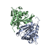
| ||||||||
|---|---|---|---|---|---|---|---|---|---|
| 1 |
| ||||||||
| Unit cell |
| ||||||||
| Noncrystallographic symmetry (NCS) | NCS oper: (Code: given Matrix: (-0.85411, 0.5099, 0.10244), Vector: |
-Components
| #1: Protein | Mass: 18732.076 Da / Num. of mol.: 2 Source method: isolated from a genetically manipulated source Source: (gene. exp.) PSEUDOMONAS AERUGINOSA (bacteria) / Strain: PAC1 (8602) / Production host: #2: Water | ChemComp-HOH / |  Mass: 18.015 Da / Num. of mol.: 180 / Source method: isolated from a natural source / Formula: H2O Mass: 18.015 Da / Num. of mol.: 180 / Source method: isolated from a natural source / Formula: H2OSequence details | IN STRAIN PAC1, RESIDUE 47 IS ALA, AS OPPOSED TO THR IN STRAIN PAO1, SEQUENCE DISCREPANCY BETWEEN ...IN STRAIN PAC1, RESIDUE 47 IS ALA, AS OPPOSED TO THR IN STRAIN PAO1, SEQUENCE DISCREPANC | |
|---|
-Experimental details
-Experiment
| Experiment | Method: X-RAY DIFFRACTION / Number of used crystals: 1 |
|---|
- Sample preparation
| Crystal | Density Matthews: 2.43 Å3/Da / Density % sol: 49 % |
|---|---|
| Crystal grow | Temperature: 291 K / Method: vapor diffusion, hanging drop / pH: 7.5 Details: HANGING DROP VAPOR DIFFUSION. RESERVOIR SOLUTION CONTAINED 100MM SODIUM CACODYLATE AT PH 6.5 OR 100MM TRIS-HCL AT PH7.5, 18-22% (W/V) PEG 10 000 AND 0.1% (W/V) NAN3. PROTEIN CONCENTRATION OF ...Details: HANGING DROP VAPOR DIFFUSION. RESERVOIR SOLUTION CONTAINED 100MM SODIUM CACODYLATE AT PH 6.5 OR 100MM TRIS-HCL AT PH7.5, 18-22% (W/V) PEG 10 000 AND 0.1% (W/V) NAN3. PROTEIN CONCENTRATION OF 10 MG/ML. DROPS KEPT AT 291K. |
-Data collection
| Diffraction | Mean temperature: 293 K |
|---|---|
| Diffraction source | Source: ROTATING ANODE / Type: ELLIOTT GX-18 / Wavelength: 1.54 |
| Detector | Type: RIGAKU RAXIS IV / Detector: IMAGE PLATE / Date: Nov 29, 2002 / Details: OSMIC MIRRORS |
| Radiation | Protocol: SINGLE WAVELENGTH / Monochromatic (M) / Laue (L): M / Scattering type: x-ray |
| Radiation wavelength | Wavelength: 1.54 Å / Relative weight: 1 |
| Reflection | Resolution: 2.35→60 Å / Num. obs: 14726 / % possible obs: 97.2 % / Observed criterion σ(I): 0 / Redundancy: 4.5 % / Biso Wilson estimate: 32.5 Å2 / Rmerge(I) obs: 0.11 / Net I/σ(I): 15.7 |
| Reflection shell | Resolution: 2.35→2.41 Å / Redundancy: 4.4 % / Rmerge(I) obs: 0.27 / Mean I/σ(I) obs: 5.8 / % possible all: 97.2 |
- Processing
| Software |
| ||||||||||||||||||||||||||||||||||||||||||||||||||||||||||||
|---|---|---|---|---|---|---|---|---|---|---|---|---|---|---|---|---|---|---|---|---|---|---|---|---|---|---|---|---|---|---|---|---|---|---|---|---|---|---|---|---|---|---|---|---|---|---|---|---|---|---|---|---|---|---|---|---|---|---|---|---|---|
| Refinement | Method to determine structure: MOLECULAR REPLACEMENT Starting model: PDB ENTRY 2J8M Resolution: 2.35→6 Å / Cross valid method: THROUGHOUT / σ(F): 0 / Stereochemistry target values: MAXIMUM LIKELIHOOD
| ||||||||||||||||||||||||||||||||||||||||||||||||||||||||||||
| Solvent computation | Bsol: 28.15 Å2 / ksol: 0.33 e/Å3 | ||||||||||||||||||||||||||||||||||||||||||||||||||||||||||||
| Displacement parameters | Biso mean: 28.7 Å2 | ||||||||||||||||||||||||||||||||||||||||||||||||||||||||||||
| Refine analyze |
| ||||||||||||||||||||||||||||||||||||||||||||||||||||||||||||
| Refinement step | Cycle: LAST / Resolution: 2.35→6 Å
| ||||||||||||||||||||||||||||||||||||||||||||||||||||||||||||
| Refine LS restraints |
|
