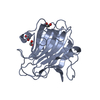
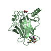
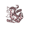
TRANSFERASE / GNAT / N-ACETYLTRANSFERASE / HYPOTHETICAL PROTEIN / PHOSPHINOTHRICIN / GCN5 FAMILY / PSEUDOMONAS AERUGINOSA
Function / homology
Function and homology information
acyltransferase activity, transferring groups other than amino-acyl groups / Transferases; Acyltransferases; Transferring groups other than aminoacyl groups Similarity search - Function
Mass: 18.015 Da / Num. of mol.: 213 / Source method: isolated from a natural source / Formula: H2O
Has protein modification
Y
Sequence details
IN STRAIN PAC1, RESIDUE 47 IS ALA, AS OPPOSED TO THR IN STRAIN PAO1 : SEQUENCE DISCREPANCY BETWEEN ...IN STRAIN PAC1, RESIDUE 47 IS ALA, AS OPPOSED TO THR IN STRAIN PAO1 : SEQUENCE DISCREPANCY BETWEEN PAC1 AND PAO1. RESIDUE 33 IS MODELLED AS ALA DUE TO DISORDER
-
Experimental details
-
Experiment
Experiment
Method: X-RAY DIFFRACTION / Number of used crystals: 1
-
Sample preparation
Crystal
Density Matthews: 2.23 Å3/Da / Density % sol: 44.39 % / Description: NONE
Crystal grow
Method: vapor diffusion, hanging drop / pH: 7.3 Details: PROTEIN WAS CRYSTALLISED USING HANGING DROP VAPOUR DIFFUSION. RESERVOIR SOLUTION CONTAINED 1ML OF 0.1M HEPES AT PH7.3, 23-27% PEG 8000 AND 0.1% AZIDE. DROP SIZE WAS 1 MICROLITRE, TO WHICH 1 ...Details: PROTEIN WAS CRYSTALLISED USING HANGING DROP VAPOUR DIFFUSION. RESERVOIR SOLUTION CONTAINED 1ML OF 0.1M HEPES AT PH7.3, 23-27% PEG 8000 AND 0.1% AZIDE. DROP SIZE WAS 1 MICROLITRE, TO WHICH 1 MICROLITRE OF PROTEIN SOLUTION AT 10 MG/ML WAS ADDED.
Protocol: SINGLE WAVELENGTH / Monochromatic (M) / Laue (L): M / Scattering type: x-ray
Radiation wavelength
Wavelength: 0.975 Å / Relative weight: 1
Reflection
Resolution: 2→70 Å / Num. obs: 13388 / % possible obs: 96.1 % / Observed criterion σ(I): 0 / Redundancy: 12.8 % / Biso Wilson estimate: 21.11 Å2 / Rmerge(I) obs: 0.1 / Net I/σ(I): 4.6
Reflection shell
Resolution: 2→2.05 Å / Redundancy: 12.7 % / Rmerge(I) obs: 0.28 / Mean I/σ(I) obs: 2.6 / % possible all: 96.1
-
Processing
Software
Name
Version
Classification
CNS
1.1
refinement
DENZO
datareduction
SCALA
datascaling
SOLVE
phasing
Refinement
Method to determine structure: SAD Starting model: NONE Resolution: 2→6 Å / Cross valid method: THROUGHOUT / σ(F): 0 / Stereochemistry target values: MAXIMUM LIKELIHOOD Details: RESIDUES A 1 AND A 2 WERE DISORDERED AND THEREFORE NOT MODELLED
Rfactor
Num. reflection
% reflection
Selection details
Rfree
0.2294
659
4.92 %
RANDOM
Rwork
0.1974
-
-
-
obs
0.1974
13388
96.1 %
-
Solvent computation
Bsol: 61.36 Å2 / ksol: 0.345 e/Å3
Displacement parameters
Biso mean: 24.04 Å2
Refine analyze
Luzzati coordinate error obs: 0.22 Å / Luzzati d res low obs: 6 Å / Luzzati sigma a obs: 0.18 Å
Refinement step
Cycle: LAST / Resolution: 2→6 Å
Protein
Nucleic acid
Ligand
Solvent
Total
Num. atoms
1303
0
35
213
1551
Refine LS restraints
Refine-ID
Type
Dev ideal
X-RAY DIFFRACTION
c_bond_d
0.019
X-RAY DIFFRACTION
c_bond_d_na
X-RAY DIFFRACTION
c_bond_d_prot
X-RAY DIFFRACTION
c_angle_d
X-RAY DIFFRACTION
c_angle_d_na
X-RAY DIFFRACTION
c_angle_d_prot
X-RAY DIFFRACTION
c_angle_deg
1.41
X-RAY DIFFRACTION
c_angle_deg_na
X-RAY DIFFRACTION
c_angle_deg_prot
X-RAY DIFFRACTION
c_dihedral_angle_d
24.68
X-RAY DIFFRACTION
c_dihedral_angle_d_na
X-RAY DIFFRACTION
c_dihedral_angle_d_prot
X-RAY DIFFRACTION
c_improper_angle_d
1.06
X-RAY DIFFRACTION
c_improper_angle_d_na
X-RAY DIFFRACTION
c_improper_angle_d_prot
X-RAY DIFFRACTION
c_mcbond_it
X-RAY DIFFRACTION
c_mcangle_it
X-RAY DIFFRACTION
c_scbond_it
X-RAY DIFFRACTION
c_scangle_it
LS refinement shell
Resolution: 2→2.05 Å / Total num. of bins used: 13 /
Rfactor
Num. reflection
Rfree
0.2509
57
Rwork
0.2656
954
+
About Yorodumi
-
News
-
Feb 9, 2022. New format data for meta-information of EMDB entries
New format data for meta-information of EMDB entries
Version 3 of the EMDB header file is now the official format.
The previous official version 1.9 will be removed from the archive.
In the structure databanks used in Yorodumi, some data are registered as the other names, "COVID-19 virus" and "2019-nCoV". Here are the details of the virus and the list of structure data.
Jan 31, 2019. EMDB accession codes are about to change! (news from PDBe EMDB page)
EMDB accession codes are about to change! (news from PDBe EMDB page)
The allocation of 4 digits for EMDB accession codes will soon come to an end. Whilst these codes will remain in use, new EMDB accession codes will include an additional digit and will expand incrementally as the available range of codes is exhausted. The current 4-digit format prefixed with “EMD-” (i.e. EMD-XXXX) will advance to a 5-digit format (i.e. EMD-XXXXX), and so on. It is currently estimated that the 4-digit codes will be depleted around Spring 2019, at which point the 5-digit format will come into force.
The EM Navigator/Yorodumi systems omit the EMD- prefix.
Related info.:Q: What is EMD? / ID/Accession-code notation in Yorodumi/EM Navigator
Yorodumi is a browser for structure data from EMDB, PDB, SASBDB, etc.
This page is also the successor to EM Navigator detail page, and also detail information page/front-end page for Omokage search.
The word "yorodu" (or yorozu) is an old Japanese word meaning "ten thousand". "mi" (miru) is to see.
Related info.:EMDB / PDB / SASBDB / Comparison of 3 databanks / Yorodumi Search / Aug 31, 2016. New EM Navigator & Yorodumi / Yorodumi Papers / Jmol/JSmol / Function and homology information / Changes in new EM Navigator and Yorodumi
 Movie
Movie Controller

 Yorodumi
 Open data
 Basic information
 Components
 Keywords
 Function and homology information
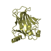
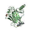
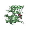
 PSEUDOMONAS AERUGINOSA (bacteria)
 X-RAY DIFFRACTION /
  Authors
 Citation
 Structure visualization
 Downloads & links
 Other downloads


 PDBj
PDBj

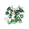
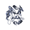
 Assembly
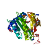

 Mass: 42.020 Da / Num. of mol.: 2 / Source method: obtained synthetically / Formula: N3
 Mass: 92.094 Da / Num. of mol.: 4 / Source method: obtained synthetically / Formula: C3H8O3
 Mass: 96.063 Da / Num. of mol.: 1 / Source method: obtained synthetically / Formula: SO4
 Mass: 18.015 Da / Num. of mol.: 213 / Source method: isolated from a natural source / Formula: H2O
 Sample preparation
 / Beamline: PX14.2 / Wavelength: 0.975
  Processing