| Entry | Database: PDB / ID: 2j7j
|
|---|
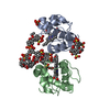
| Title | Invariance of the zinc finger module: a comparison of the free structure with those in nucleic-acid complexes |
|---|
 Components Components | TRANSCRIPTION FACTOR IIIA |
|---|
 Keywords Keywords | TRANSCRIPTION / ZINC FINGER MODULE / ALTERNATIVE INITIATION / NUCLEAR PROTEIN / PHOSPHORYLATION / HYDROPHOBIC CORE / ZINC / RNA-BINDING / ZINC-FINGER / DNA-BINDING / TRANSCRIPTION REGULATION / POLYMORPHISM / METAL-BINDING |
|---|
| Function / homology |  Function and homology information Function and homology information
: / TFIIIA, beta-beta-alpha zinc finger / : / Classic Zinc Finger / Double Stranded RNA Binding Domain / Zinc finger, C2H2 type / zinc finger / Zinc finger C2H2 type domain profile. / Zinc finger C2H2 superfamily / Zinc finger C2H2 type domain signature. ...: / TFIIIA, beta-beta-alpha zinc finger / : / Classic Zinc Finger / Double Stranded RNA Binding Domain / Zinc finger, C2H2 type / zinc finger / Zinc finger C2H2 type domain profile. / Zinc finger C2H2 superfamily / Zinc finger C2H2 type domain signature. / Zinc finger C2H2-type / 2-Layer Sandwich / Alpha BetaSimilarity search - Domain/homology |
|---|
| Biological species |  XENOPUS LAEVIS (African clawed frog) XENOPUS LAEVIS (African clawed frog) |
|---|
| Method |  X-RAY DIFFRACTION / SYNCHROTRON / MAD / Resolution: 1.65 Å X-RAY DIFFRACTION / SYNCHROTRON / MAD / Resolution: 1.65 Å |
|---|
 Authors Authors | Lu, D. / Klug, A. |
|---|
 Citation Citation | Journal: Proteins / Year: 2007
Title: Invariance of the Zinc Finger Module: A Comparison of the Free Structure with Those in Nucleic-Acid Complexes.
Authors: Lu, D. / Klug, A. |
|---|
| History | | Deposition | Oct 10, 2006 | Deposition site: PDBE / Processing site: PDBE |
|---|
| Revision 1.0 | Oct 9, 2007 | Provider: repository / Type: Initial release |
|---|
| Revision 1.1 | Oct 10, 2012 | Group: Database references / Derived calculations ...Database references / Derived calculations / Non-polymer description / Other / Version format compliance |
|---|
| Revision 1.2 | May 8, 2024 | Group: Data collection / Database references / Derived calculations
Category: chem_comp_atom / chem_comp_bond ...chem_comp_atom / chem_comp_bond / database_2 / pdbx_struct_conn_angle / struct_conn / struct_site
Item: _database_2.pdbx_DOI / _database_2.pdbx_database_accession ..._database_2.pdbx_DOI / _database_2.pdbx_database_accession / _pdbx_struct_conn_angle.ptnr1_auth_comp_id / _pdbx_struct_conn_angle.ptnr1_auth_seq_id / _pdbx_struct_conn_angle.ptnr1_label_atom_id / _pdbx_struct_conn_angle.ptnr1_label_comp_id / _pdbx_struct_conn_angle.ptnr1_label_seq_id / _pdbx_struct_conn_angle.ptnr3_auth_comp_id / _pdbx_struct_conn_angle.ptnr3_auth_seq_id / _pdbx_struct_conn_angle.ptnr3_label_atom_id / _pdbx_struct_conn_angle.ptnr3_label_comp_id / _pdbx_struct_conn_angle.ptnr3_label_seq_id / _pdbx_struct_conn_angle.value / _struct_conn.pdbx_dist_value / _struct_conn.ptnr1_auth_comp_id / _struct_conn.ptnr1_auth_seq_id / _struct_conn.ptnr1_label_asym_id / _struct_conn.ptnr1_label_atom_id / _struct_conn.ptnr1_label_comp_id / _struct_conn.ptnr1_label_seq_id / _struct_conn.ptnr2_auth_comp_id / _struct_conn.ptnr2_auth_seq_id / _struct_conn.ptnr2_label_asym_id / _struct_conn.ptnr2_label_atom_id / _struct_conn.ptnr2_label_comp_id / _struct_conn.ptnr2_label_seq_id / _struct_site.pdbx_auth_asym_id / _struct_site.pdbx_auth_comp_id / _struct_site.pdbx_auth_seq_id |
|---|
|
|---|
 Movie
Movie Controller

 Yorodumi
 Open data
 Basic information
 Structure visualization
 Downloads & links
 Other downloads


 PDBj
PDBj


 Assembly


 Mass: 65.409 Da / Num. of mol.: 3 / Source method: obtained synthetically / Formula: Zn
 Mass: 96.063 Da / Num. of mol.: 1 / Source method: obtained synthetically / Formula: SO4
 Mass: 18.015 Da / Num. of mol.: 128 / Source method: isolated from a natural source / Formula: H2O
 Sample preparation
 / Beamline: ID29 / Wavelength: 0.9791, 1.2824, 1.2828, 1.2651
 Processing