 Movie
Movie Controller

[English] 日本語
 Yorodumi
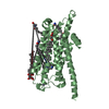
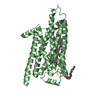
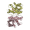
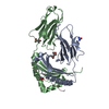
- PDB-1un6: THE CRYSTAL STRUCTURE OF A ZINC FINGER - RNA COMPLEX REVEALS TWO ... -
+ Open data

- Basic information
| Entry | Database: PDB / ID: 1un6 | ||||||
|---|---|---|---|---|---|---|---|
| Title | THE CRYSTAL STRUCTURE OF A ZINC FINGER - RNA COMPLEX REVEALS TWO MODES OF MOLECULAR RECOGNITION | ||||||
 Components Components |
| ||||||
 Keywords Keywords | RNA-BINDING PROTEIN/RNA / COMPLEX(ZINC FINGER-RNA) / TFIIIA / 5S RIBOSOMAL RNA / ZINC FINGER / RNA-PROTEIN COMPLEX / X. LAEVIS / TRANSCRIPTION REGULATION / RNA-BINDING / DNA-BINDING / NUCLEAR PROTEIN / RNA-BINDING PROTEIN-RNA complex | ||||||
| Function / homology |  Function and homology information Function and homology informationribosomal large subunit biogenesis / DNA binding / RNA binding / zinc ion binding / nucleus Similarity search - Function | ||||||
| Biological species | |||||||
| Method |  X-RAY DIFFRACTION / SYNCHROTRON / MAD / Resolution: 3.1 Å X-RAY DIFFRACTION / SYNCHROTRON / MAD / Resolution: 3.1 Å | ||||||
 Authors Authors | Lu, D. / Searles, M.A. / Klug, A. | ||||||
 Citation Citation | Journal: Nature / Year: 2003 Title: Crystal Structure of a Zinc-Finger-RNA Complex Reveals Two Modes of Molecular Recognition Authors: Lu, D. / Searles, M.A. / Klug, A. #1: Journal: J.Mol.Biol. / Year: 2000 Title: The Role of the Central Zinc Fingers of Transcription Factor Iiia in Binding to 5S RNA Authors: Searles, M.A. / Lu, D. / Klug, A. | ||||||
| History |
| ||||||
| Remark 650 | HELIX DETERMINATION METHOD: AUTHOR PROVIDED. | ||||||
| Remark 700 | SHEET DETERMINATION METHOD: AUTHOR PROVIDED. |
- Structure visualization
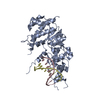
| Structure viewer | Molecule: MolmilJmol/JSmol |
|---|
- Downloads & links
-Download
| PDBx/mmCIF format | 1un6.cif.gz | 132 KB | Display | PDBx/mmCIF format |
|---|---|---|---|---|
| PDB format | pdb1un6.ent.gz | 98 KB | Display | PDB format |
| PDBx/mmJSON format | 1un6.json.gz | Tree view | PDBx/mmJSON format | |
| Others |  Other downloads Other downloads |
-Validation report
| Arichive directory | https://data.pdbj.org/pub/pdb/validation_reports/un/1un6ftp://data.pdbj.org/pub/pdb/validation_reports/un/1un6 | HTTPS FTP |
|---|
-Related structure data
| Related structure data | |
|---|---|
| Similar structure data |


-Links
 PDBj
PDBj


- Assembly
| Deposited unit | 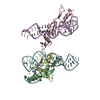
| ||||||||
|---|---|---|---|---|---|---|---|---|---|
| 1 | 
| ||||||||
| 2 | 
| ||||||||
| Unit cell |
| ||||||||
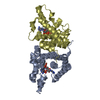
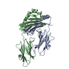
| Details | THE ENTRY CONTAINS TWO COPIES OF THE RNA- PROTEIN COMPLEXAND AN EXTRA PROTEIN WITH CHAIN IDENTIFIER D. THE TWOCOPIES OF RNA ARE IN CHAIN IDENTIFIERS E AND F, AND THE TWOCOPIES OF THE PROTEIN IN THE COMPLEXES ARE IN THE CHAINIDENTIFIERS B AND C.THE DIMER DESCRIBED IN REMARK 350 DOES NOT REFLECTA BIOLOGICALLY FUNCTIONAL DIMER. |
-Components
| #1: Protein | Mass: 10299.835 Da / Num. of mol.: 3 Fragment: FINGERS 4,5 AND 6, RESIDUES 127 - 212 UNDER SWISSPROT NUMBERING FOR SOMATIC TFIIIA Source method: isolated from a genetically manipulated source Source: (gene. exp.)  #2: RNA chain | Mass: 19708.777 Da / Num. of mol.: 2 Fragment: CENTRAL REGION, NUCLEOTIDES 4 - 15,64 -82,94-115, PLUS TWO TETRALOOPS JOINING 15 - 64 AND 82 -94 RESPECTIVELY Source method: isolated from a natural source / Details: IN VITRO TRANSCRIPTION TO PRODUCE THE RNA / Source: (natural) #3: Chemical | ChemComp-ZN /   Mass: 65.409 Da / Num. of mol.: 8 / Source method: obtained synthetically / Formula: Zn Mass: 65.409 Da / Num. of mol.: 8 / Source method: obtained synthetically / Formula: Zn#4: Chemical | ChemComp-MG /   Mass: 24.305 Da / Num. of mol.: 13 / Source method: obtained synthetically / Formula: Mg Mass: 24.305 Da / Num. of mol.: 13 / Source method: obtained synthetically / Formula: Mg#5: Water | ChemComp-HOH / |  Mass: 18.015 Da / Num. of mol.: 16 / Source method: isolated from a natural source / Formula: H2O Mass: 18.015 Da / Num. of mol.: 16 / Source method: isolated from a natural source / Formula: H2OCompound details | ACTS BOTH AS A POSITIVE TRANSCRIPTION FACTOR FOR 5S RNA GENES AND A SPECIFIC RNA BINDING PROTEIN ...ACTS BOTH AS A POSITIVE TRANSCRIPT | |
|---|
-Experimental details
-Experiment
| Experiment | Method: X-RAY DIFFRACTION / Number of used crystals: 2 |
|---|
- Sample preparation
| Crystal | Density Matthews: 3.12 Å3/Da / Density % sol: 50 % | ||||||||||||||||||||||||||||||||||||||||||||||||||||||||
|---|---|---|---|---|---|---|---|---|---|---|---|---|---|---|---|---|---|---|---|---|---|---|---|---|---|---|---|---|---|---|---|---|---|---|---|---|---|---|---|---|---|---|---|---|---|---|---|---|---|---|---|---|---|---|---|---|---|
| Crystal grow | pH: 5.6 Details: 20% PEG 8000, 200MM KCL, 5MM MGCL2, 50MM MES, PH 5.6, 3MM DTT, 0.3MM ZNSO4 | ||||||||||||||||||||||||||||||||||||||||||||||||||||||||
| Crystal grow | *PLUS Temperature: 4 ℃ / pH: 5.6 / Method: vapor diffusion, hanging drop | ||||||||||||||||||||||||||||||||||||||||||||||||||||||||
| Components of the solutions | *PLUS
|
-Data collection
| Diffraction |
| ||||||||||||||||||
|---|---|---|---|---|---|---|---|---|---|---|---|---|---|---|---|---|---|---|---|
| Diffraction source |
| ||||||||||||||||||
| Detector |
| ||||||||||||||||||
| Radiation |
| ||||||||||||||||||
| Radiation wavelength |
| ||||||||||||||||||
| Reflection | Resolution: 3.1→35.2 Å / Num. obs: 15267 / % possible obs: 97.9 % / Observed criterion σ(I): 2 / Redundancy: 3.8 % / Rmerge(I) obs: 0.051 / Net I/σ(I): 7.9 | ||||||||||||||||||
| Reflection shell | Resolution: 3.1→3.27 Å / Redundancy: 3.9 % / Rmerge(I) obs: 0.315 / Mean I/σ(I) obs: 2 / % possible all: 97.5 | ||||||||||||||||||
| Reflection | *PLUS Highest resolution: 3.1 Å / Rmerge(I) obs: 0.051 |


- Processing
| Software |
| ||||||||||||||||||||||||||||||||||||||||||||||||||||||||||||||||||||||||||||||||
|---|---|---|---|---|---|---|---|---|---|---|---|---|---|---|---|---|---|---|---|---|---|---|---|---|---|---|---|---|---|---|---|---|---|---|---|---|---|---|---|---|---|---|---|---|---|---|---|---|---|---|---|---|---|---|---|---|---|---|---|---|---|---|---|---|---|---|---|---|---|---|---|---|---|---|---|---|---|---|---|---|---|
| Refinement | Method to determine structure: MAD / Resolution: 3.1→35.19 Å / Rfactor Rfree error: 0.009 / Data cutoff high absF: 2782617.75 / Isotropic thermal model: RESTRAINED / Cross valid method: THROUGHOUT / σ(F): 0 Details: REFMAC5 WAS USED TO REACH R=0.2 AND RFREE=0.3, THEN THE MODEL WAS REFINED IN CNS.
| ||||||||||||||||||||||||||||||||||||||||||||||||||||||||||||||||||||||||||||||||
| Solvent computation | Solvent model: FLAT MODEL / Bsol: 35.8942 Å2 / ksol: 0.272189 e/Å3 | ||||||||||||||||||||||||||||||||||||||||||||||||||||||||||||||||||||||||||||||||
| Displacement parameters | Biso mean: 92 Å2
| ||||||||||||||||||||||||||||||||||||||||||||||||||||||||||||||||||||||||||||||||
| Refine analyze |
| ||||||||||||||||||||||||||||||||||||||||||||||||||||||||||||||||||||||||||||||||
| Refinement step | Cycle: LAST / Resolution: 3.1→35.19 Å
| ||||||||||||||||||||||||||||||||||||||||||||||||||||||||||||||||||||||||||||||||
| Refine LS restraints |
| ||||||||||||||||||||||||||||||||||||||||||||||||||||||||||||||||||||||||||||||||
| LS refinement shell | Resolution: 3.1→3.29 Å / Rfactor Rfree error: 0.031 / Total num. of bins used: 6
| ||||||||||||||||||||||||||||||||||||||||||||||||||||||||||||||||||||||||||||||||
| Xplor file |
| ||||||||||||||||||||||||||||||||||||||||||||||||||||||||||||||||||||||||||||||||
| Refinement | *PLUS Highest resolution: 3.1 Å / Rfactor Rfree: 0.256 / Rfactor Rwork: 0.215 | ||||||||||||||||||||||||||||||||||||||||||||||||||||||||||||||||||||||||||||||||
| Solvent computation | *PLUS | ||||||||||||||||||||||||||||||||||||||||||||||||||||||||||||||||||||||||||||||||
| Displacement parameters | *PLUS | ||||||||||||||||||||||||||||||||||||||||||||||||||||||||||||||||||||||||||||||||
| Refine LS restraints | *PLUS
|
