 Movie
Movie Controller

[English] 日本語
 Yorodumi
- PDB-3dv2: Crystal Structure of nicotinic acid mononucleotide adenylyltransf... -
+ Open data

- Basic information
| Entry | Database: PDB / ID: 3dv2 | ||||||
|---|---|---|---|---|---|---|---|
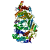
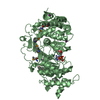
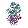
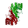
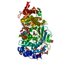
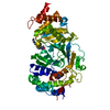
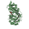
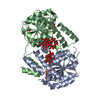
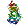
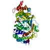
| Title | Crystal Structure of nicotinic acid mononucleotide adenylyltransferase from Bacillus anthracis | ||||||
 Components Components | Nicotinate (Nicotinamide) nucleotide adenylyltransferase | ||||||
 Keywords Keywords | TRANSFERASE / alpha and beta protein / Rossmann fold / NAD / Nucleotidyltransferase / Pyridine nucleotide biosynthesis | ||||||
| Function / homology | HUPs / Rossmann fold / 3-Layer(aba) Sandwich / Alpha Beta / :  Function and homology information Function and homology information | ||||||
| Biological species |  | ||||||
| Method |  X-RAY DIFFRACTION / SYNCHROTRON / MOLECULAR REPLACEMENT / molecular replacement / Resolution: 2.3 Å X-RAY DIFFRACTION / SYNCHROTRON / MOLECULAR REPLACEMENT / molecular replacement / Resolution: 2.3 Å | ||||||
 Authors Authors | Lu, S. / Smith, C.D. / Yang, Z. / Pruett, P.S. / Nagy, L. / McCombs, D.P. / DeLucas, L.J. / Brouillette, W.J. / Brouillette, C.G. | ||||||
 Citation Citation | Journal: ACTA CRYSTALLOGR.,SECT.F / Year: 2008 Title: Structure of nicotinic acid mononucleotide adenylyltransferase from Bacillus anthracis. Authors: Lu, S. / Smith, C.D. / Yang, Z. / Pruett, P.S. / Nagy, L. / McCombs, D. / Delucas, L.J. / Brouillette, W.J. / Brouillette, C.G. | ||||||
| History |
|
- Structure visualization
| Structure viewer | Molecule: MolmilJmol/JSmol |
|---|
- Downloads & links
-Download
| PDBx/mmCIF format | 3dv2.cif.gz | 165.1 KB | Display | PDBx/mmCIF format |
|---|---|---|---|---|
| PDB format | pdb3dv2.ent.gz | 131.5 KB | Display | PDB format |
| PDBx/mmJSON format | 3dv2.json.gz | Tree view | PDBx/mmJSON format | |
| Others |  Other downloads Other downloads |
-Validation report
| Arichive directory | https://data.pdbj.org/pub/pdb/validation_reports/dv/3dv2ftp://data.pdbj.org/pub/pdb/validation_reports/dv/3dv2 | HTTPS FTP |
|---|
-Related structure data
| Related structure data |  1kamS S: Starting model for refinement |
|---|---|
| Similar structure data |
-Links
 PDBj
PDBj- Assembly
| Deposited unit | 
| ||||||||
|---|---|---|---|---|---|---|---|---|---|
| 1 | 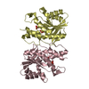
| ||||||||
| 2 | 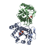
| ||||||||
| Unit cell |
|
-Components
| #1: Protein | Mass: 23379.869 Da / Num. of mol.: 4 Source method: isolated from a genetically manipulated source Source: (gene. exp.) References: UniProt: Q6HT60, nicotinate-nucleotide adenylyltransferase #2: Chemical | ChemComp-SO4 /   Mass: 96.063 Da / Num. of mol.: 8 / Source method: obtained synthetically / Formula: SO4 Mass: 96.063 Da / Num. of mol.: 8 / Source method: obtained synthetically / Formula: SO4#3: Water | ChemComp-HOH / |  Mass: 18.015 Da / Num. of mol.: 129 / Source method: isolated from a natural source / Formula: H2O Mass: 18.015 Da / Num. of mol.: 129 / Source method: isolated from a natural source / Formula: H2O |
|---|
-Experimental details
-Experiment
| Experiment | Method: X-RAY DIFFRACTION / Number of used crystals: 1 |
|---|
- Sample preparation
| Crystal | Density Matthews: 2.33 Å3/Da / Density % sol: 47.3 % |
|---|---|
| Crystal grow | Temperature: 295 K / Method: vapor diffusion, hanging drop / pH: 7 Details: 2.1M ammonium sulfate, 0.1M HEPES, 2% PEG400, 10mM magnesium chloride, pH 7.0, VAPOR DIFFUSION, HANGING DROP, temperature 295K |
-Data collection
| Diffraction | Mean temperature: 100 K |
|---|---|
| Diffraction source | Source: SYNCHROTRON / Site: APS  / Beamline: 22-ID / Wavelength: 1 Å / Beamline: 22-ID / Wavelength: 1 Å |
| Detector | Type: MARMOSAIC 300 mm CCD / Detector: CCD / Date: Oct 12, 2007 |
| Radiation | Monochromator: GRAPHITE / Protocol: SINGLE WAVELENGTH / Monochromatic (M) / Laue (L): M / Scattering type: x-ray |
| Radiation wavelength | Wavelength: 1 Å / Relative weight: 1 |
| Reflection | Resolution: 2.3→25.15 Å / Num. all: 38428 / Num. obs: 37472 / % possible obs: 97.5 % / Observed criterion σ(F): 1 / Observed criterion σ(I): 1 / Redundancy: 3.7 % / Rmerge(I) obs: 0.059 / Rsym value: 0.059 / Net I/σ(I): 10.5 |
| Reflection shell | Resolution: 2.3→2.38 Å / Redundancy: 3.3 % / Rmerge(I) obs: 0.285 / Mean I/σ(I) obs: 3.2 / Rsym value: 0.285 / % possible all: 88.9 |
-Phasing
| Phasing | Method: molecular replacement |
|---|
- Processing
| Software |
| ||||||||||||||||||||||||||||||||||||||||||||||||||||||||||||||||||||||||||||||||
|---|---|---|---|---|---|---|---|---|---|---|---|---|---|---|---|---|---|---|---|---|---|---|---|---|---|---|---|---|---|---|---|---|---|---|---|---|---|---|---|---|---|---|---|---|---|---|---|---|---|---|---|---|---|---|---|---|---|---|---|---|---|---|---|---|---|---|---|---|---|---|---|---|---|---|---|---|---|---|---|---|---|
| Refinement | Method to determine structure: MOLECULAR REPLACEMENT Starting model: PDB ENTRY 1KAM Resolution: 2.3→25.15 Å / Rfactor Rfree error: 0.006 / Occupancy max: 1 / Occupancy min: 1 / Data cutoff high absF: 566579 / Data cutoff low absF: 0 / Isotropic thermal model: RESTRAINED / Cross valid method: THROUGHOUT / σ(F): 0 / Details: BULK SOLVENT MODEL USED
| ||||||||||||||||||||||||||||||||||||||||||||||||||||||||||||||||||||||||||||||||
| Solvent computation | Solvent model: FLAT MODEL / Bsol: 42.93 Å2 / ksol: 0.35 e/Å3 | ||||||||||||||||||||||||||||||||||||||||||||||||||||||||||||||||||||||||||||||||
| Displacement parameters | Biso mean: 44.44 Å2
| ||||||||||||||||||||||||||||||||||||||||||||||||||||||||||||||||||||||||||||||||
| Refine analyze |
| ||||||||||||||||||||||||||||||||||||||||||||||||||||||||||||||||||||||||||||||||
| Refinement step | Cycle: LAST / Resolution: 2.3→25.15 Å
| ||||||||||||||||||||||||||||||||||||||||||||||||||||||||||||||||||||||||||||||||
| Refine LS restraints |
| ||||||||||||||||||||||||||||||||||||||||||||||||||||||||||||||||||||||||||||||||
| LS refinement shell | Resolution: 2.3→2.44 Å / Rfactor Rfree error: 0.022 / Total num. of bins used: 6
| ||||||||||||||||||||||||||||||||||||||||||||||||||||||||||||||||||||||||||||||||
| Xplor file |
|
