 Movie
Movie Controller


+ Open data

- Basic information
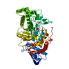
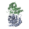
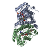
| Entry | Database: PDB / ID: 6tuw | ||||||
|---|---|---|---|---|---|---|---|
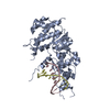
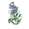
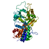
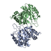
| Title | human XPG-DNA, Complex 1 | ||||||
 Components Components |
| ||||||
 Keywords Keywords | DNA BINDING PROTEIN / XPG nuclease domain bound to DNA | ||||||
| Function / homology |  Function and homology information Function and homology informationnucleotide-excision repair complex / base-excision repair, AP site formation / bubble DNA binding / hydrolase activity, acting on ester bonds / response to UV-C / RNA polymerase II complex binding / response to UV / transcription-coupled nucleotide-excision repair / nucleotide-excision repair / DNA endonuclease activity ...nucleotide-excision repair complex / base-excision repair, AP site formation / bubble DNA binding / hydrolase activity, acting on ester bonds / response to UV-C / RNA polymerase II complex binding / response to UV / transcription-coupled nucleotide-excision repair / nucleotide-excision repair / DNA endonuclease activity / double-strand break repair via homologous recombination / Dual Incision in GG-NER / enzyme activator activity / Formation of Incision Complex in GG-NER / Dual incision in TC-NER / single-stranded DNA binding / chromosome / double-stranded DNA binding / endonuclease activity / damaged DNA binding / Hydrolases; Acting on ester bonds / negative regulation of apoptotic process / protein-containing complex binding / protein homodimerization activity / protein-containing complex / nucleoplasm / metal ion binding / nucleus Similarity search - Function | ||||||
| Biological species |  Homo sapiens (human) Homo sapiens (human)unidentified (others) | ||||||
| Method |  X-RAY DIFFRACTION / SYNCHROTRON / MOLECULAR REPLACEMENT / Resolution: 3.5 Å X-RAY DIFFRACTION / SYNCHROTRON / MOLECULAR REPLACEMENT / Resolution: 3.5 Å | ||||||
 Authors Authors | Ruiz, F.M. / Fernandez-Tornero, C. | ||||||
 Citation Citation | Journal: Nucleic Acids Res. / Year: 2020 Title: The crystal structure of human XPG, the xeroderma pigmentosum group G endonuclease, provides insight into nucleotide excision DNA repair. Authors: Gonzalez-Corrochano, R. / Ruiz, F.M. / Taylor, N.M.I. / Huecas, S. / Drakulic, S. / Spinola-Amilibia, M. / Fernandez-Tornero, C. | ||||||
| History |
|
- Structure visualization
| Structure viewer | Molecule: MolmilJmol/JSmol |
|---|
- Downloads & links
-Download
| PDBx/mmCIF format | 6tuw.cif.gz | 107.5 KB | Display | PDBx/mmCIF format |
|---|---|---|---|---|
| PDB format | pdb6tuw.ent.gz | 65.1 KB | Display | PDB format |
| PDBx/mmJSON format | 6tuw.json.gz | Tree view | PDBx/mmJSON format | |
| Others |  Other downloads Other downloads |
-Validation report
| Arichive directory | https://data.pdbj.org/pub/pdb/validation_reports/tu/6tuwftp://data.pdbj.org/pub/pdb/validation_reports/tu/6tuw | HTTPS FTP |
|---|
-Related structure data
| Related structure data |  6turSC  6tusC  6tuxC S: Starting model for refinement C: citing same article ( |
|---|---|
| Similar structure data |


-Links
 PDBj
PDBj


- Assembly
| Deposited unit | 
| ||||||||||||
|---|---|---|---|---|---|---|---|---|---|---|---|---|---|
| 1 |
| ||||||||||||
| Unit cell |
|
-Components
| #1: Protein | Mass: 40789.887 Da / Num. of mol.: 1 Source method: isolated from a genetically manipulated source Source: (gene. exp.) Homo sapiens (human) / Gene: ERCC5, ERCM2, XPG, XPGC / Production host:  References: UniProt: P28715, Hydrolases; Acting on ester bonds |
|---|---|
| #2: DNA chain | Mass: 3364.208 Da / Num. of mol.: 1 / Source method: obtained synthetically / Source: (synth.) unidentified (others) |
| #3: DNA chain | Mass: 3358.211 Da / Num. of mol.: 1 / Source method: obtained synthetically / Source: (synth.) unidentified (others) |
-Experimental details
-Experiment
| Experiment | Method: X-RAY DIFFRACTION / Number of used crystals: 1 |
|---|
- Sample preparation
| Crystal | Density Matthews: 2.62 Å3/Da / Density % sol: 53.05 % |
|---|---|
| Crystal grow | Temperature: 295 K / Method: vapor diffusion, sitting drop / Details: 5% PEG 3350, 50 mM Na Citrate pH 4.0 |
-Data collection
| Diffraction | Mean temperature: 100 K / Serial crystal experiment: N |
|---|---|
| Diffraction source | Source: SYNCHROTRON / Site: ESRF  / Beamline: ID29 / Wavelength: 0.9795 Å / Beamline: ID29 / Wavelength: 0.9795 Å |
| Detector | Type: DECTRIS PILATUS 6M-F / Detector: PIXEL / Date: Feb 1, 2015 |
| Radiation | Protocol: SINGLE WAVELENGTH / Monochromatic (M) / Laue (L): M / Scattering type: x-ray |
| Radiation wavelength | Wavelength: 0.9795 Å / Relative weight: 1 |
| Reflection | Resolution: 3.5→45.79 Å / Num. obs: 6627 / % possible obs: 99.8 % / Redundancy: 15.9 % / Biso Wilson estimate: 145.35 Å2 / CC1/2: 0.998 / Net I/σ(I): 11.1 |
| Reflection shell | Resolution: 3.5→3.625 Å / Num. unique obs: 1536 / CC1/2: 0.827 |
- Processing
| Software |
| ||||||||||||||||||||||||
|---|---|---|---|---|---|---|---|---|---|---|---|---|---|---|---|---|---|---|---|---|---|---|---|---|---|
| Refinement | Method to determine structure: MOLECULAR REPLACEMENT Starting model: 6TUR Resolution: 3.5→45.79 Å / SU ML: 0.3407 / Cross valid method: FREE R-VALUE / σ(F): 1.34 / Phase error: 39.6614
| ||||||||||||||||||||||||
| Solvent computation | Shrinkage radii: 0.9 Å / VDW probe radii: 1.11 Å | ||||||||||||||||||||||||
| Displacement parameters | Biso mean: 212.3 Å2 | ||||||||||||||||||||||||
| Refinement step | Cycle: LAST / Resolution: 3.5→45.79 Å
| ||||||||||||||||||||||||
| Refine LS restraints |
| ||||||||||||||||||||||||
| LS refinement shell |
|
