Synthesis of bile acids and bile salts via 24-hydroxycholesterol / chlordecone reductase activity / steroid dehydrogenase activity, acting on the CH-OH group of donors, NAD or NADP as acceptor / RA biosynthesis pathway / Retinoid metabolism and transport / 3(or 17)alpha-hydroxysteroid dehydrogenase / 17-beta-hydroxysteroid dehydrogenase (NADP+) activity / trans-1,2-dihydrobenzene-1,2-diol dehydrogenase activity / Synthesis of bile acids and bile salts via 7alpha-hydroxycholesterol / Synthesis of bile acids and bile salts via 27-hydroxycholesterol ...Synthesis of bile acids and bile salts via 24-hydroxycholesterol / chlordecone reductase activity / steroid dehydrogenase activity, acting on the CH-OH group of donors, NAD or NADP as acceptor / RA biosynthesis pathway / Retinoid metabolism and transport / 3(or 17)alpha-hydroxysteroid dehydrogenase / 17-beta-hydroxysteroid dehydrogenase (NADP+) activity / trans-1,2-dihydrobenzene-1,2-diol dehydrogenase activity / Synthesis of bile acids and bile salts via 7alpha-hydroxycholesterol / Synthesis of bile acids and bile salts via 27-hydroxycholesterol / Synthesis of Prostaglandins (PG) and Thromboxanes (TX) / Prednisone ADME / prostaglandin D2 11-ketoreductase activity / ketoreductase activity / 5-alpha-androstane-3-beta,17-beta-diol dehydrogenase (NADP+) activity / 15-hydroxyprostaglandin-D dehydrogenase (NADP+) activity / Delta4-3-oxosteroid 5beta-reductase activity / : / 3-alpha-hydroxysteroid 3-dehydrogenase [NAD(P)+] activity / androsterone dehydrogenase [NAD(P)+] activity / ketosteroid monooxygenase activity / progesterone metabolic process / steroid biosynthetic process / carboxylic acid binding / all-trans-retinol dehydrogenase (NAD+) activity / alcohol dehydrogenase (NADP+) activity / lithocholic acid binding / prostaglandin H2 endoperoxidase reductase activity / Oxidoreductases; Acting on the CH-OH group of donors; With NAD+ or NADP+ as acceptor / bile acid binding / daunorubicin metabolic process / doxorubicin metabolic process / retinal dehydrogenase (NAD+) activity / aldose reductase (NADPH) activity / oxidoreductase activity, acting on NAD(P)H, quinone or similar compound as acceptor / prostaglandin metabolic process / NADP+ binding / steroid metabolic process / NADPH binding / steroid binding / nucleus / cytoplasm / cytosol Similarity search - Function
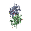
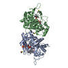
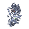
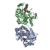
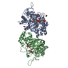
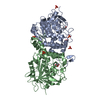
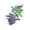
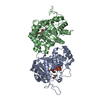
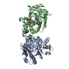
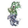
Aldo-keto reductase family 1 member C / NADP-dependent oxidoreductase domain / Aldo/keto reductase family signature 2. / Aldo/keto reductase, conserved site / Aldo-keto reductase / NADP-dependent oxidoreductase domain / Aldo/keto reductase family / NADP-dependent oxidoreductase domain superfamily / TIM Barrel / Alpha-Beta Barrel / Alpha Beta Similarity search - Domain/homology
BETA-MERCAPTOETHANOL / Chem-FFA / NADP NICOTINAMIDE-ADENINE-DINUCLEOTIDE PHOSPHATE / Aldo-keto reductase family 1 member C21 Similarity search - Component
Biological species
Mus musculus (house mouse)
Method
X-RAY DIFFRACTION / MOLECULAR REPLACEMENT / Resolution: 1.9 Å
In the structure databanks used in Yorodumi, some data are registered as the other names, "COVID-19 virus" and "2019-nCoV". Here are the details of the virus and the list of structure data.
Jan 31, 2019. EMDB accession codes are about to change! (news from PDBe EMDB page)
EMDB accession codes are about to change! (news from PDBe EMDB page)
The allocation of 4 digits for EMDB accession codes will soon come to an end. Whilst these codes will remain in use, new EMDB accession codes will include an additional digit and will expand incrementally as the available range of codes is exhausted. The current 4-digit format prefixed with “EMD-” (i.e. EMD-XXXX) will advance to a 5-digit format (i.e. EMD-XXXXX), and so on. It is currently estimated that the 4-digit codes will be depleted around Spring 2019, at which point the 5-digit format will come into force.
The EM Navigator/Yorodumi systems omit the EMD- prefix.
Related info.:Q: What is EMD? / ID/Accession-code notation in Yorodumi/EM Navigator
Yorodumi is a browser for structure data from EMDB, PDB, SASBDB, etc.
This page is also the successor to EM Navigator detail page, and also detail information page/front-end page for Omokage search.
The word "yorodu" (or yorozu) is an old Japanese word meaning "ten thousand". "mi" (miru) is to see.
Related info.:EMDB / PDB / SASBDB / Comparison of 3 databanks / Yorodumi Search / Aug 31, 2016. New EM Navigator & Yorodumi / Yorodumi Papers / Jmol/JSmol / Function and homology information / Changes in new EM Navigator and Yorodumi
 Movie
Movie Controller

 Yorodumi
 Open data
 Basic information
 Components
 Keywords
 Function and homology information
 X-RAY DIFFRACTION /
  Authors
 Citation
 Structure visualization
 Downloads & links
 Other downloads
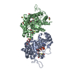

 PDBj
PDBj


 Assembly
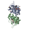


 Mass: 743.405 Da / Num. of mol.: 2 / Mutation: K31A / Source method: obtained synthetically / Formula: C21H28N7O17P3
 Mass: 288.424 Da / Num. of mol.: 2 / Source method: obtained synthetically / Formula: C19H28O2 / Comment: inhibitor, antagonist, hormone*YM
 Mass: 78.133 Da / Num. of mol.: 4 / Source method: obtained synthetically / Formula: C2H6OS
 Mass: 62.068 Da / Num. of mol.: 3 / Source method: obtained synthetically / Formula: C2H6O2
 Sample preparation
 Processing