 Movie
Movie Controller

[English] 日本語
 Yorodumi
- PDB-2ijo: Crystal Structure of the West Nile virus NS2B-NS3 protease comple... -
+ Open data

- Basic information
| Entry | Database: PDB / ID: 2ijo | ||||||
|---|---|---|---|---|---|---|---|
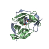
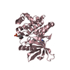
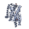
| Title | Crystal Structure of the West Nile virus NS2B-NS3 protease complexed with bovine pancreatic trypsin inhibitor | ||||||
 Components Components |
| ||||||
 Keywords Keywords | HYDROLASE/HYDROLASE INHIBITOR / WEST NILE VIRUS / PROTEASE / APROTININ / BPTI / NS2B / NS3 / FLAVIVIRUS / serine protease / HYDROLASE-HYDROLASE INHIBITOR COMPLEX | ||||||
| Function / homology |  Function and homology information Function and homology informationRNA 5'-cap (guanine-N7)-methylation / DNA/DNA annealing activity / symbiont-mediated suppression of host JAK-STAT cascade via inhibition of JAK1 activity / RNA strand annealing activity / RNA stabilization / sulfate binding / symbiont-mediated suppression of host apoptosis / negative regulation of platelet aggregation / RNA folding chaperone / zymogen binding ...RNA 5'-cap (guanine-N7)-methylation / DNA/DNA annealing activity / symbiont-mediated suppression of host JAK-STAT cascade via inhibition of JAK1 activity / RNA strand annealing activity / RNA stabilization / sulfate binding / symbiont-mediated suppression of host apoptosis / negative regulation of platelet aggregation / RNA folding chaperone / zymogen binding / potassium channel inhibitor activity / molecular function inhibitor activity / negative regulation of thrombin-activated receptor signaling pathway / flavivirin / symbiont-mediated suppression of host JAK-STAT cascade via inhibition of host TYK2 activity / serine protease inhibitor complex / positive regulation of viral genome replication / symbiont-mediated suppression of host JAK-STAT cascade via inhibition of STAT2 activity / symbiont-mediated suppression of host JAK-STAT cascade via inhibition of STAT1 activity / serine-type endopeptidase inhibitor activity / protein-DNA complex / viral capsid / ribonucleoside triphosphate phosphatase activity / peptidase activity / nucleoside-triphosphate phosphatase / double-stranded RNA binding / protease binding / clathrin-dependent endocytosis of virus by host cell / mRNA (guanine-N7)-methyltransferase / methyltransferase cap1 / methyltransferase cap1 activity / mRNA 5'-cap (guanine-N7-)-methyltransferase activity / RNA helicase activity / protein dimerization activity / symbiont-mediated suppression of host innate immune response / host cell perinuclear region of cytoplasm / host cell endoplasmic reticulum membrane / RNA helicase / symbiont-mediated suppression of host type I interferon-mediated signaling pathway / ribonucleoprotein complex / serine-type endopeptidase activity / symbiont-mediated activation of host autophagy / RNA-directed RNA polymerase / viral RNA genome replication / RNA-directed RNA polymerase activity / fusion of virus membrane with host endosome membrane / viral envelope / calcium ion binding / symbiont entry into host cell / virion attachment to host cell / host cell nucleus / virion membrane / structural molecule activity / ATP hydrolysis activity / proteolysis / : / DNA binding / RNA binding / extracellular region / ATP binding / metal ion binding Similarity search - Function | ||||||
| Biological species |  West Nile virus West Nile virus | ||||||
| Method |  X-RAY DIFFRACTION / SYNCHROTRON / MOLECULAR REPLACEMENT / Resolution: 2.3 Å X-RAY DIFFRACTION / SYNCHROTRON / MOLECULAR REPLACEMENT / Resolution: 2.3 Å | ||||||
 Authors Authors | Aleshin, A.E. / Shiryaev, S.A. / Strongin, A.Y. / Liddington, R.C. | ||||||
 Citation Citation | Journal: Protein Sci. / Year: 2007 Title: Structural evidence for regulation and specificity of flaviviral proteases and evolution of the Flaviviridae fold. Authors: Aleshin, A.E. / Shiryaev, S.A. / Strongin, A.Y. / Liddington, R.C. | ||||||
| History |
| ||||||
| Remark 400 | COMPOUND THE PROTEINS NS2B (CHAIN A) AND NS3 (CHAIN B) ARE CONNECTED THROUGH A LINKER AGGGGSGGGG. | ||||||
| Remark 999 | SEQUENCE THIS IS THE SEQUENCE OF THE LINKER BETWEEN CHAINS A AND B. RESIDUE NUMBERS 1 THROUGH 5 IN ... SEQUENCE THIS IS THE SEQUENCE OF THE LINKER BETWEEN CHAINS A AND B. RESIDUE NUMBERS 1 THROUGH 5 IN COORDINATES FOR CHAIN B ARE ARBITRARY, SINCE ONLY THESE FIVE RESIDUES WERE VISIBLE IN ELECTRON DENSITY. THE SEQUENCE ALIGNMENT OF THESE FIVE RESIDUES IS ALSO ARBITRARY AND IS BASED ONLY ON THE DISTANCE CRITERIA TO THE NEXT VISIBLE RESIDUES. |
- Structure visualization
| Structure viewer | Molecule: MolmilJmol/JSmol |
|---|
- Downloads & links
-Download
| PDBx/mmCIF format | 2ijo.cif.gz | 67.2 KB | Display | PDBx/mmCIF format |
|---|---|---|---|---|
| PDB format | pdb2ijo.ent.gz | 48.3 KB | Display | PDB format |
| PDBx/mmJSON format | 2ijo.json.gz | Tree view | PDBx/mmJSON format | |
| Others |  Other downloads Other downloads |
-Validation report
| Arichive directory | https://data.pdbj.org/pub/pdb/validation_reports/ij/2ijoftp://data.pdbj.org/pub/pdb/validation_reports/ij/2ijo | HTTPS FTP |
|---|
-Related structure data
| Related structure data | 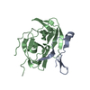 2ggvSC S: Starting model for refinement C: citing same article ( |
|---|---|
| Similar structure data |


-Links
 PDBj
PDBj


- Assembly
| Deposited unit | 
| ||||||||
|---|---|---|---|---|---|---|---|---|---|
| 1 |
| ||||||||
| Unit cell |
| ||||||||
| Details | NS2B cofactor and NS3 protease domain form heterodimer |
-Components
| #1: Protein | Mass: 6078.417 Da / Num. of mol.: 1 / Fragment: NS2B cofactor domain Source method: isolated from a genetically manipulated source Source: (gene. exp.) West Nile virus / Genus: Flavivirus / Plasmid: pET101 / Production host:  References: UniProt: Q203W3, UniProt: P06935*PLUS, flavivirin |
|---|---|
| #2: Protein | Mass: 20906.584 Da / Num. of mol.: 1 / Fragment: NS3 protease domain / Mutation: K104R Source method: isolated from a genetically manipulated source Source: (gene. exp.) West Nile virus / Genus: Flavivirus / Plasmid: pET101 / Production host: References: UniProt: Q203W3, UniProt: P06935*PLUS, flavivirin |
| #3: Protein | Mass: 6527.568 Da / Num. of mol.: 1 / Source method: isolated from a natural source / Source: (natural) |
| #4: Water | ChemComp-HOH /  Mass: 18.015 Da / Num. of mol.: 89 / Source method: isolated from a natural source / Formula: H2O Mass: 18.015 Da / Num. of mol.: 89 / Source method: isolated from a natural source / Formula: H2O |
| Has protein modification | Y |
-Experimental details
-Experiment
| Experiment | Method: X-RAY DIFFRACTION / Number of used crystals: 1 |
|---|
- Sample preparation
| Crystal | Density Matthews: 1.97 Å3/Da / Density % sol: 37.5 % |
|---|---|
| Crystal grow | Temperature: 298 K / Method: vapor diffusion, hanging drop / pH: 8.5 Details: 20% PEG 8000, 0.1M TRIS-HCL, 0.2M SODIUM CHLORIDE, pH 8.5, VAPOR DIFFUSION, HANGING DROP, temperature 298K |
-Data collection
| Diffraction | Mean temperature: 170 K |
|---|---|
| Diffraction source | Source: SYNCHROTRON / Site: ALS  / Beamline: 12.3.1 / Wavelength: 1 Å / Beamline: 12.3.1 / Wavelength: 1 Å |
| Detector | Type: ADSC QUANTUM 4 / Detector: CCD / Date: Feb 28, 2006 |
| Radiation | Protocol: SINGLE WAVELENGTH / Monochromatic (M) / Laue (L): M / Scattering type: x-ray |
| Radiation wavelength | Wavelength: 1 Å / Relative weight: 1 |
| Reflection | Resolution: 2.3→40 Å / Num. all: 11411 / Num. obs: 11411 / % possible obs: 93 % / Observed criterion σ(F): 0 / Observed criterion σ(I): 0 / Redundancy: 7.4 % / Biso Wilson estimate: 47 Å2 / Rmerge(I) obs: 0.069 / Net I/σ(I): 24 |
| Reflection shell | Resolution: 2.3→2.45 Å / Redundancy: 3.7 % / Rmerge(I) obs: 0.33 / Mean I/σ(I) obs: 2.9 / % possible all: 70 |
- Processing
| Software |
| ||||||||||||||||||||||||||||
|---|---|---|---|---|---|---|---|---|---|---|---|---|---|---|---|---|---|---|---|---|---|---|---|---|---|---|---|---|---|
| Refinement | Method to determine structure: MOLECULAR REPLACEMENT Starting model: PDB entry 2GGV Resolution: 2.3→40 Å / Isotropic thermal model: anisotropic / Cross valid method: THROUGHOUT / σ(F): 0 / Stereochemistry target values: Engh & Huber
| ||||||||||||||||||||||||||||
| Solvent computation | Bsol: 42.735 Å2 | ||||||||||||||||||||||||||||
| Displacement parameters | Biso mean: 44.735 Å2
| ||||||||||||||||||||||||||||
| Refinement step | Cycle: LAST / Resolution: 2.3→40 Å
| ||||||||||||||||||||||||||||
| Refine LS restraints |
| ||||||||||||||||||||||||||||
| Xplor file |
|
