- PDB-2iaa: Crystal Structure of an Electron Transfer Complex Between Aromati... -
+
Open data
ID or keywords:
Loading...
-
Basic information
Entry
Database: PDB / ID: 2iaa
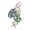
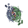
Title
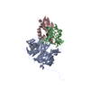
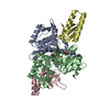
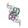
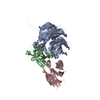
Crystal Structure of an Electron Transfer Complex Between Aromatic Amine Dephydrogenase and Azurin from Alcaligenes Faecalis (Form 2)
Components
(Aromatic Amine Dehydrogenase) x 2
Azurin
Keywords
OXIDOREDUCTASE/electron transport / Quinoprotein / tryptophan tryptophylquinone / cupredoxin / electron transfer / OXIDOREDUCTASE-electron transport COMPLEX
Function / homology
Function and homology information
aralkylamine dehydrogenase (azurin) / aralkylamine dehydrogenase (azurin) activity / aliphatic amine dehydrogenase activity / amine metabolic process / periplasmic space / electron transfer activity / copper ion binding Similarity search - Function
Amine dehydrogenase heavy chain / Methylamine/Aralkylamine dehydrogenase light chain, C-terminal domain / Amine dehydrogenase light chain / Methylamine/Aralkylamine dehydrogenase light chain superfamily / Methylamine dehydrogenase, L chain / Methylamine dehydrogenase heavy chain (MADH) / Electron Transport Ethylamine Dehydrogenase / Methylamine/Aralkylamine dehydrogenase light chain / : / Azurin ...Amine dehydrogenase heavy chain / Methylamine/Aralkylamine dehydrogenase light chain, C-terminal domain / Amine dehydrogenase light chain / Methylamine/Aralkylamine dehydrogenase light chain superfamily / Methylamine dehydrogenase, L chain / Methylamine dehydrogenase heavy chain (MADH) / Electron Transport Ethylamine Dehydrogenase / Methylamine/Aralkylamine dehydrogenase light chain / : / Azurin / Quinoprotein amine dehydrogenase, beta chain-like / : / Blue (type 1) copper domain / Copper binding proteins, plastocyanin/azurin family / Blue (type 1) copper protein, binding site / Type-1 copper (blue) proteins signature. / Cupredoxins - blue copper proteins / Twin arginine translocation (Tat) signal profile. / Twin-arginine translocation pathway, signal sequence / YVTN repeat-like/Quinoprotein amine dehydrogenase / Cupredoxin / 7 Propeller / Methylamine Dehydrogenase; Chain H / WD40/YVTN repeat-like-containing domain superfamily / Immunoglobulin-like / Sandwich / Mainly Beta Similarity search - Domain/homology
COPPER (II) ION / Azurin / Aralkylamine dehydrogenase light chain / Aralkylamine dehydrogenase heavy chain Similarity search - Component
Biological species
Alcaligenes faecalis (bacteria)
Method
X-RAY DIFFRACTION / SYNCHROTRON / MAD / Resolution: 1.95 Å
SEQUENCE THE SEQUENCE OF AROMATIC AMINE DEHYDROGENASE (CHAINS A,D,B,E) ARE NOT AVAILABLE AT UNP ...SEQUENCE THE SEQUENCE OF AROMATIC AMINE DEHYDROGENASE (CHAINS A,D,B,E) ARE NOT AVAILABLE AT UNP SEQUENCE DATABASE AT THE TIME OF PROCESSING.
Remark 300
BIOMOLECULE: 1,2 THIS ENTRY CONTAINS THE CRYSTALLOGRAPHIC ASYMMETRIC UNIT WHICH CONSISTS OF 5 ...BIOMOLECULE: 1,2 THIS ENTRY CONTAINS THE CRYSTALLOGRAPHIC ASYMMETRIC UNIT WHICH CONSISTS OF 5 CHAIN(S). AUTHOR STATES THAT THE ASYMMETRIC UNIT CONTAINS ONE ALPHA-BETA-GAMMA HETEROTRIMER WHICH IS HALF THE BIOLOGICAL UNIT FOR A BINARY COMPLEX AND ONE ALPHA-BETA HETERODIMER WHICH IS NOT THE BIOLOGICAL ELECTRON TRANSFER BIOLOGICAL UNIT.
In the structure databanks used in Yorodumi, some data are registered as the other names, "COVID-19 virus" and "2019-nCoV". Here are the details of the virus and the list of structure data.
Jan 31, 2019. EMDB accession codes are about to change! (news from PDBe EMDB page)
EMDB accession codes are about to change! (news from PDBe EMDB page)
The allocation of 4 digits for EMDB accession codes will soon come to an end. Whilst these codes will remain in use, new EMDB accession codes will include an additional digit and will expand incrementally as the available range of codes is exhausted. The current 4-digit format prefixed with “EMD-” (i.e. EMD-XXXX) will advance to a 5-digit format (i.e. EMD-XXXXX), and so on. It is currently estimated that the 4-digit codes will be depleted around Spring 2019, at which point the 5-digit format will come into force.
The EM Navigator/Yorodumi systems omit the EMD- prefix.
Related info.:Q: What is EMD? / ID/Accession-code notation in Yorodumi/EM Navigator
Yorodumi is a browser for structure data from EMDB, PDB, SASBDB, etc.
This page is also the successor to EM Navigator detail page, and also detail information page/front-end page for Omokage search.
The word "yorodu" (or yorozu) is an old Japanese word meaning "ten thousand". "mi" (miru) is to see.
Related info.:EMDB / PDB / SASBDB / Comparison of 3 databanks / Yorodumi Search / Aug 31, 2016. New EM Navigator & Yorodumi / Yorodumi Papers / Jmol/JSmol / Function and homology information / Changes in new EM Navigator and Yorodumi
 Movie
Movie Controller

 Yorodumi
 Open data
 Basic information
 Components
 Keywords
 Function and homology information
 Alcaligenes faecalis (bacteria)
 X-RAY DIFFRACTION /
  Authors
 Citation
 Structure visualization
 Downloads & links
 Other downloads
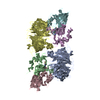


 PDBj
PDBj

 Assembly
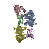

 Mass: 63.546 Da / Num. of mol.: 1 / Source method: obtained synthetically / Formula: Cu
 Mass: 18.015 Da / Num. of mol.: 989 / Source method: isolated from a natural source / Formula: H2O
 Sample preparation
 / Beamline: 19-ID / Wavelength: 1.381, 1.445
 Processing