 Movie
Movie Controller

[English] 日本語
 Yorodumi
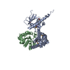
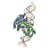
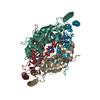
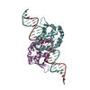
- PDB-2h47: Crystal Structure of an Electron Transfer Complex Between Aromati... -
+ Open data

- Basic information
| Entry | Database: PDB / ID: 2h47 | ||||||
|---|---|---|---|---|---|---|---|
| Title | Crystal Structure of an Electron Transfer Complex Between Aromatic Amine Dephydrogenase and Azurin from Alcaligenes Faecalis (Form 1) | ||||||
 Components Components |
| ||||||
 Keywords Keywords | OXIDOREDUCTASE/electron transport / Quinoprotein / tryptophan tryptophylquinone / cupredoxin / electron transfer / OXIDOREDUCTASE-electron transport COMPLEX | ||||||
| Function / homology |  Function and homology information Function and homology informationaralkylamine dehydrogenase (azurin) / aralkylamine dehydrogenase (azurin) activity / aliphatic amine dehydrogenase activity / amine metabolic process / periplasmic space / electron transfer activity / copper ion binding Similarity search - Function | ||||||
| Biological species |  Alcaligenes faecalis (bacteria) Alcaligenes faecalis (bacteria) | ||||||
| Method |  X-RAY DIFFRACTION / MOLECULAR REPLACEMENT / Resolution: 2.6 Å X-RAY DIFFRACTION / MOLECULAR REPLACEMENT / Resolution: 2.6 Å | ||||||
 Authors Authors | Sukumar, N. / Chen, Z. / Leys, D. / Scrutton, N.S. / Ferrati, D. / Merli, A. / Rossi, G.L. / Bellamy, H.D. / Chistoserdov, A. / Davidson, V.L. / Mathews, F.S. | ||||||
 Citation Citation | Journal: Biochemistry / Year: 2006 Title: Crystal Structure of an Electron Transfer Complex between Aromatic Amine Dehydrogenase and Azurin from Alcaligenes faecalis. Authors: Sukumar, N. / Chen, Z. / Ferrari, D. / Merli, A. / Rossi, G.L. / Bellamy, H.D. / Chistoserdov, A. / Davidson, V.L. / Mathews, F.S. #1: Journal: Science / Year: 2006Title: Atomic Description of an Enzyme Reaction Dominated by Proton Tunneling Authors: Masgrau, L. / Roujeinikova, A. / Johannissen, L.O. / Hothi, P. / Basran, J. / Ranaghan, K.E. / Mulholland, A.J. / Sutcliffe, M.J. / Scrutton, N.S. / Leys, D. | ||||||
| History |
| ||||||
| Remark 999 | SEQUENCE THE SEQUENCE OF AROMATIC AMINE DEHYDROGENASE (CHAINS A,D,F,H,B,E,G,I) ARE NOT AVAILABLE AT ...SEQUENCE THE SEQUENCE OF AROMATIC AMINE DEHYDROGENASE (CHAINS A,D,F,H,B,E,G,I) ARE NOT AVAILABLE AT UNP SEQUENCE DATABASE AT THE TIME OF PROCESSING. | ||||||
| Remark 300 | BIOMOLECULE: 1,2,3,4 THIS ENTRY CONTAINS THE CRYSTALLOGRAPHIC ASYMMETRIC UNIT WHICH CONSISTS OF 9 ...BIOMOLECULE: 1,2,3,4 THIS ENTRY CONTAINS THE CRYSTALLOGRAPHIC ASYMMETRIC UNIT WHICH CONSISTS OF 9 CHAIN(S). AUTHOR STATES THAT THE ASYMMETRIC UNIT CONTAINS ONE ALPHA-BETA-GAMMA HETEROTRIMER WHICH IS HALF THE BIOLOGICAL UNIT FOR A BINARY COMPLEX AND AND THREE ALPHA-BETA HETERODIMERS, TWO OF WHICH FORM UNCOMPLEXED HETEROTETRAMER AND THE THIRD ASSOCIATES WITH THE HETEROTRIMER COMPLEX. |
- Structure visualization
| Structure viewer | Molecule: MolmilJmol/JSmol |
|---|
- Downloads & links
-Download
| PDBx/mmCIF format | 2h47.cif.gz | 418.7 KB | Display | PDBx/mmCIF format |
|---|---|---|---|---|
| PDB format | pdb2h47.ent.gz | 339.7 KB | Display | PDB format |
| PDBx/mmJSON format | 2h47.json.gz | Tree view | PDBx/mmJSON format | |
| Others |  Other downloads Other downloads |
-Validation report
| Arichive directory | https://data.pdbj.org/pub/pdb/validation_reports/h4/2h47ftp://data.pdbj.org/pub/pdb/validation_reports/h4/2h47 | HTTPS FTP |
|---|
-Related structure data
| Related structure data |  2h3xC  2iaaC  2ah1S  2h3y S: Starting model for refinement C: citing same article ( |
|---|---|
| Similar structure data |

-Links
 PDBj
PDBj

- Assembly
| Deposited unit | 
| ||||||||
|---|---|---|---|---|---|---|---|---|---|
| 1 | 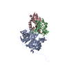
| ||||||||
| 2 | 
| ||||||||
| 3 | 
| ||||||||
| 4 | 
| ||||||||
| 5 | 
| ||||||||
| 6 | 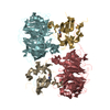
| ||||||||
| Unit cell |
|
-Components
| #1: Protein | Mass: 42978.754 Da / Num. of mol.: 4 / Source method: isolated from a natural source / Source: (natural) Alcaligenes faecalis (bacteria) / Strain: IFO 14479 / References: UniProt: P84888, EC: 1.4.99.4#2: Protein | Mass: 14516.898 Da / Num. of mol.: 4 / Source method: isolated from a natural source / Source: (natural) Alcaligenes faecalis (bacteria) / Strain: IFO 14479 / References: UniProt: P84887, EC: 1.4.99.4#3: Protein | | Mass: 13711.415 Da / Num. of mol.: 1 / Source method: isolated from a natural source / Source: (natural) Alcaligenes faecalis (bacteria) / Strain: IFO 14479 / References: UniProt: P00281#4: Chemical | ChemComp-CU / |   Mass: 63.546 Da / Num. of mol.: 1 / Source method: obtained synthetically / Formula: Cu Mass: 63.546 Da / Num. of mol.: 1 / Source method: obtained synthetically / Formula: Cu#5: Water | ChemComp-HOH / |  Mass: 18.015 Da / Num. of mol.: 747 / Source method: isolated from a natural source / Formula: H2O Mass: 18.015 Da / Num. of mol.: 747 / Source method: isolated from a natural source / Formula: H2O |
|---|
-Experimental details
-Experiment
| Experiment | Method: X-RAY DIFFRACTION / Number of used crystals: 1 |
|---|
- Sample preparation
| Crystal | Density Matthews: 2.37 Å3/Da / Density % sol: 48.01 % |
|---|---|
| Crystal grow | Temperature: 295 K / Method: vapor diffusion, sitting drop / pH: 8.5 Details: 23-28% PEG 4000, 0.1M Tris and 0.2M sodium acetate, pH 8.5, VAPOR DIFFUSION, SITTING DROP, temperature 295K |
-Data collection
| Diffraction | Mean temperature: 100 K |
|---|---|
| Diffraction source | Source: ROTATING ANODE / Type: RIGAKU RU200 / Wavelength: 1.5418 Å |
| Detector | Type: RIGAKU RAXIS IV / Detector: IMAGE PLATE / Date: Mar 26, 2001 |
| Radiation | Monochromator: GRAPHITE / Protocol: SINGLE WAVELENGTH / Monochromatic (M) / Laue (L): M / Scattering type: x-ray |
| Radiation wavelength | Wavelength: 1.5418 Å / Relative weight: 1 |
| Reflection | Resolution: 2.6→40 Å / Num. all: 72744 / Num. obs: 50339 / % possible obs: 69.2 % / Observed criterion σ(F): 0 / Observed criterion σ(I): 0 / Redundancy: 3.4 % / Biso Wilson estimate: 35.2 Å2 / Rmerge(I) obs: 0.058 / Net I/σ(I): 17.8 |
| Reflection shell | Resolution: 2.6→2.69 Å / Redundancy: 3.5 % / Rmerge(I) obs: 0.118 / Mean I/σ(I) obs: 5.9 / Num. unique all: 2818 / % possible all: 39.2 |
- Processing
| Software |
| |||||||||||||||||||||||||||||||||||||||||||||||||
|---|---|---|---|---|---|---|---|---|---|---|---|---|---|---|---|---|---|---|---|---|---|---|---|---|---|---|---|---|---|---|---|---|---|---|---|---|---|---|---|---|---|---|---|---|---|---|---|---|---|---|
| Refinement | Method to determine structure: MOLECULAR REPLACEMENT Starting model: PDB ENTRY 2AH1 Resolution: 2.6→34.01 Å / Rfactor Rfree error: 0.003 / Data cutoff high absF: 605127.55 / Data cutoff low absF: 0 / Isotropic thermal model: Isotropic / Cross valid method: THROUGHOUT / σ(F): 0 / Stereochemistry target values: Engh & Huber
| |||||||||||||||||||||||||||||||||||||||||||||||||
| Solvent computation | Solvent model: FLAT MODEL / Bsol: 29.2491 Å2 / ksol: 0.343646 e/Å3 | |||||||||||||||||||||||||||||||||||||||||||||||||
| Displacement parameters | Biso mean: 28.1 Å2
| |||||||||||||||||||||||||||||||||||||||||||||||||
| Refine analyze |
| |||||||||||||||||||||||||||||||||||||||||||||||||
| Refinement step | Cycle: LAST / Resolution: 2.6→34.01 Å
| |||||||||||||||||||||||||||||||||||||||||||||||||
| Refine LS restraints |
| |||||||||||||||||||||||||||||||||||||||||||||||||
| LS refinement shell | Refine-ID: X-RAY DIFFRACTION / Total num. of bins used: 6
|
