- PDB-2h3x: Crystal Structure of an Electron Transfer Complex Between Aromati... -
+
データを開く
IDまたはキーワード:
読み込み中...
-
基本情報
登録情報
データベース: PDB / ID: 2h3x
タイトル
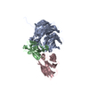
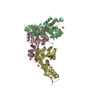
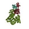
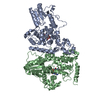
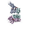
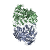
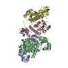
Crystal Structure of an Electron Transfer Complex Between Aromatic Amine Dehydrogenase and Azurin from Alcaligenes Faecalis (Form 3)
要素
(Aromatic Amine Dehydrogenase) x 2
Azurin
キーワード
OXIDOREDUCTASE/electron transport / Quinoprotein / tryptophan tryptophylquinone / cupredoxin / electron transfer / OXIDOREDUCTASE-electron transport COMPLEX
機能・相同性
機能・相同性情報
aralkylamine dehydrogenase (azurin) / aralkylamine dehydrogenase (azurin) activity / aliphatic amine dehydrogenase activity / amine metabolic process / periplasmic space / electron transfer activity / copper ion binding 類似検索 - 分子機能
Amine dehydrogenase heavy chain / Methylamine/Aralkylamine dehydrogenase light chain, C-terminal domain / Amine dehydrogenase light chain / Methylamine/Aralkylamine dehydrogenase light chain superfamily / Methylamine dehydrogenase, L chain / Methylamine dehydrogenase heavy chain (MADH) / Electron Transport Ethylamine Dehydrogenase / Methylamine/Aralkylamine dehydrogenase light chain / : / Azurin ...Amine dehydrogenase heavy chain / Methylamine/Aralkylamine dehydrogenase light chain, C-terminal domain / Amine dehydrogenase light chain / Methylamine/Aralkylamine dehydrogenase light chain superfamily / Methylamine dehydrogenase, L chain / Methylamine dehydrogenase heavy chain (MADH) / Electron Transport Ethylamine Dehydrogenase / Methylamine/Aralkylamine dehydrogenase light chain / : / Azurin / Quinoprotein amine dehydrogenase, beta chain-like / : / Blue (type 1) copper domain / Copper binding proteins, plastocyanin/azurin family / Blue (type 1) copper protein, binding site / Type-1 copper (blue) proteins signature. / Cupredoxins - blue copper proteins / Twin arginine translocation (Tat) signal profile. / Twin-arginine translocation pathway, signal sequence / YVTN repeat-like/Quinoprotein amine dehydrogenase / Cupredoxin / 7 Propeller / Methylamine Dehydrogenase; Chain H / WD40/YVTN repeat-like-containing domain superfamily / Immunoglobulin-like / Sandwich / Mainly Beta 類似検索 - ドメイン・相同性
COPPER (II) ION / Azurin / Aralkylamine dehydrogenase light chain / Aralkylamine dehydrogenase heavy chain 類似検索 - 構成要素
SEQUENCE THE SEQUENCE OF AROMATIC AMINE DEHYDROGENASE (CHAINS A,D,B,E) ARE NOT AVAILABLE AT UNP ...SEQUENCE THE SEQUENCE OF AROMATIC AMINE DEHYDROGENASE (CHAINS A,D,B,E) ARE NOT AVAILABLE AT UNP SEQUENCE DATABASE AT THE TIME OF PROCESSING.
 ムービー
ムービー コントローラー


 データを開く
 基本情報
基本情報 要素
要素 キーワード
キーワード 機能・相同性情報
 Alcaligenes faecalis (バクテリア)
 X線回折 /
  データ登録者
 引用
引用 構造の表示
構造の表示 ダウンロードとリンク
 その他のダウンロード
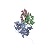


 PDBj
PDBj

 集合体
集合体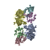

 分子量: 63.546 Da / 分子数: 2 / 由来タイプ: 合成 / 式: Cu
 分子量: 18.015 Da / 分子数: 651 / 由来タイプ: 天然 / 式: H2O
 試料調製
試料調製 / ビームライン: 8-BM / 波長: 1.3753 Å
 解析
解析