 Movie
Movie Controller

[English] 日本語
 Yorodumi
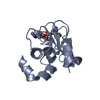
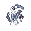
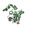
- PDB-2i3h: Structure of an ML-IAP/XIAP chimera bound to a 4-mer peptide (AVPW) -
+ Open data

- Basic information
| Entry | Database: PDB / ID: 2i3h | ||||||
|---|---|---|---|---|---|---|---|
| Title | Structure of an ML-IAP/XIAP chimera bound to a 4-mer peptide (AVPW) | ||||||
 Components Components |
| ||||||
 Keywords Keywords | INHIBITOR/APOPTOSIS / ZINC BINDING / PEPTIDE COMPLEX / APOPTOSIS INHIBITION / PEPTIDOMIMETIC / SMALL MOLECULE / DRUG DESIGN / INHIBITOR-APOPTOSIS COMPLEX | ||||||
| Function / homology |  Function and homology information Function and homology informationregulation of natural killer cell apoptotic process / cysteine-type endopeptidase inhibitor activity involved in apoptotic process / Regulation of MITF-M-dependent genes involved in apoptosis / lens development in camera-type eye / cysteine-type endopeptidase inhibitor activity / negative regulation of tumor necrosis factor-mediated signaling pathway / positive regulation of protein ubiquitination / RING-type E3 ubiquitin transferase / positive regulation of JNK cascade / ubiquitin-protein transferase activity ...regulation of natural killer cell apoptotic process / cysteine-type endopeptidase inhibitor activity involved in apoptotic process / Regulation of MITF-M-dependent genes involved in apoptosis / lens development in camera-type eye / cysteine-type endopeptidase inhibitor activity / negative regulation of tumor necrosis factor-mediated signaling pathway / positive regulation of protein ubiquitination / RING-type E3 ubiquitin transferase / positive regulation of JNK cascade / ubiquitin-protein transferase activity / ubiquitin protein ligase activity / regulation of cell population proliferation / regulation of apoptotic process / regulation of cell cycle / protein ubiquitination / apoptotic process / centrosome / negative regulation of apoptotic process / enzyme binding / Golgi apparatus / zinc ion binding / nucleoplasm / nucleus / cytoplasm / cytosol Similarity search - Function | ||||||
| Biological species |  Homo sapiens (human) Homo sapiens (human) | ||||||
| Method |  X-RAY DIFFRACTION / SYNCHROTRON / FOURIER SYNTHESIS / Resolution: 1.62 Å X-RAY DIFFRACTION / SYNCHROTRON / FOURIER SYNTHESIS / Resolution: 1.62 Å | ||||||
 Authors Authors | Fairbrother, W.J. / Franklin, M.C. | ||||||
 Citation Citation | Journal: Acs Chem.Biol. / Year: 2006 Title: Design, synthesis, and biological activity of a potent Smac mimetic that sensitizes cancer cells to apoptosis by antagonizing IAPs. Authors: Zobel, K. / Wang, L. / Varfolomeev, E. / Franklin, M.C. / Elliott, L.O. / Wallweber, H.J. / Okawa, D.C. / Flygare, J.A. / Vucic, D. / Fairbrother, W.J. / Deshayes, K. #1: Journal: Biochem.J. / Year: 2005Title: Engineering ML-IAP to produce an extraordinarily potent caspase-9 inhibitor: implications for Smac-dependent anti-apoptotic activity of ML-IAP Authors: Vucic, D. / Franklin, M.C. / Wallweber, H.J. / Das, K. / Eckelman, B.P. / Shin, H. / Elliott, L.O. / Deshayes, K. / Salvesen, G.S. / Fairbrother, W.J. #2: Journal: Biochemistry / Year: 2003Title: Structure and Function Analysis of Peptide Antagonists of Melanoma Inhibitor of Apoptosis (ML-IAP) Authors: Franklin, M.C. / Kadkhodayan, S. / Ackerly, H. / Alexandru, D. / Distefano, M.D. / Elliott, L.O. / Flygare, J.A. / Vucic, D. / Deshayes, K. / Fairbrother, W.J. | ||||||
| History |
| ||||||
| Remark 999 | SEQUENCE For entity 1 (chains A and B) residues 150, 160-168, and 172 replaced with XIAP-BIR3 homologues. |
- Structure visualization
| Structure viewer | Molecule: MolmilJmol/JSmol |
|---|
- Downloads & links
-Download
| PDBx/mmCIF format | 2i3h.cif.gz | 102.6 KB | Display | PDBx/mmCIF format |
|---|---|---|---|---|
| PDB format | pdb2i3h.ent.gz | 77.2 KB | Display | PDB format |
| PDBx/mmJSON format | 2i3h.json.gz | Tree view | PDBx/mmJSON format | |
| Others |  Other downloads Other downloads |
-Validation report
| Arichive directory | https://data.pdbj.org/pub/pdb/validation_reports/i3/2i3hftp://data.pdbj.org/pub/pdb/validation_reports/i3/2i3h | HTTPS FTP |
|---|
-Related structure data


-Links
 PDBj
PDBj


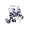
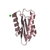
- Assembly
| Deposited unit | 
| ||||||||
|---|---|---|---|---|---|---|---|---|---|
| 1 | 
| ||||||||
| 2 | 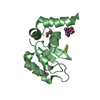
| ||||||||
| 3 | 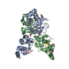
| ||||||||
| 4 | 
| ||||||||
| 5 | 
| ||||||||
| 6 | 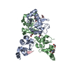
| ||||||||
| 7 | 
| ||||||||
| Unit cell |
|
-Components
-Protein / Protein/peptide , 2 types, 4 molecules ABCD
| #1: Protein | Mass: 14977.567 Da / Num. of mol.: 2 / Fragment: ML-IAP residues 63-172 Mutation: RESIDUES 150, 160-168, AND 172 REPLACED WITH XIAP-BIR3 HOMOLOGUES Source method: isolated from a genetically manipulated source Source: (gene. exp.) Homo sapiens (human) / Gene: BIRC7 / Plasmid: pET15b / Species (production host): Escherichia coli / Production host:  #2: Protein/peptide | Mass: 471.548 Da / Num. of mol.: 2 / Source method: obtained synthetically / Details: Chemically synthesized peptide |
|---|
-Non-polymers , 5 types, 239 molecules 


| #3: Chemical |  Mass: 65.409 Da / Num. of mol.: 2 / Source method: obtained synthetically / Formula: Zn Mass: 65.409 Da / Num. of mol.: 2 / Source method: obtained synthetically / Formula: Zn#4: Chemical | ChemComp-LI / |  Mass: 6.941 Da / Num. of mol.: 1 / Source method: obtained synthetically / Formula: Li Mass: 6.941 Da / Num. of mol.: 1 / Source method: obtained synthetically / Formula: Li#5: Chemical | ChemComp-BTB / |  Mass: 209.240 Da / Num. of mol.: 1 / Source method: obtained synthetically / Formula: C8H19NO5 / Comment: pH buffer*YM Mass: 209.240 Da / Num. of mol.: 1 / Source method: obtained synthetically / Formula: C8H19NO5 / Comment: pH buffer*YM#6: Chemical |  Mass: 62.068 Da / Num. of mol.: 2 / Source method: obtained synthetically / Formula: C2H6O2 Mass: 62.068 Da / Num. of mol.: 2 / Source method: obtained synthetically / Formula: C2H6O2#7: Water | ChemComp-HOH / | Mass: 18.015 Da / Num. of mol.: 233 / Source method: isolated from a natural source / Formula: H2O |
|---|
-Experimental details
-Experiment
| Experiment | Method: X-RAY DIFFRACTION / Number of used crystals: 1 |
|---|
- Sample preparation
| Crystal | Density Matthews: 2.29 Å3/Da / Density % sol: 46.26 % |
|---|---|
| Crystal grow | Temperature: 298 K / Method: vapor diffusion, hanging drop / pH: 6.5 Details: Lithium sulfate, PEG 3350, Bis-tris, pH 6.5, VAPOR DIFFUSION, HANGING DROP, temperature 298K |
-Data collection
| Diffraction | Mean temperature: 100 K |
|---|---|
| Diffraction source | Source: SYNCHROTRON / Site: APS  / Beamline: 19-BM / Wavelength: 1.0332 Å / Beamline: 19-BM / Wavelength: 1.0332 Å |
| Detector | Type: SBC / Detector: CCD / Date: Aug 15, 2003 |
| Radiation | Monochromator: Rosenbaum-Rock double-crystal monochromator: Water cooled; sagitally focusing 2nd crystal, Rosenbaum-Rock vertical focusing mirror Protocol: SINGLE WAVELENGTH / Monochromatic (M) / Laue (L): M / Scattering type: x-ray |
| Radiation wavelength | Wavelength: 1.0332 Å / Relative weight: 1 |
| Reflection | Resolution: 1.62→50 Å / Num. all: 37115 / Num. obs: 32349 / % possible obs: 87.2 % / Observed criterion σ(F): 0 / Observed criterion σ(I): -3 / Redundancy: 5.9 % / Biso Wilson estimate: 16 Å2 / Rmerge(I) obs: 0.068 / Net I/σ(I): 20.9 |
| Reflection shell | Resolution: 1.62→1.68 Å / Redundancy: 0.7 % / Rmerge(I) obs: 0.406 / Mean I/σ(I) obs: 1.7 / Num. unique all: 1397 / % possible all: 38.5 |
- Processing
| Software |
| ||||||||||||||||||||||||||||||||||||||||||||||||||||||||||||||||||||||||||||||||||||||||||||||||||||||||||||||||||||||||||||||||||||||||||||||||||||||
|---|---|---|---|---|---|---|---|---|---|---|---|---|---|---|---|---|---|---|---|---|---|---|---|---|---|---|---|---|---|---|---|---|---|---|---|---|---|---|---|---|---|---|---|---|---|---|---|---|---|---|---|---|---|---|---|---|---|---|---|---|---|---|---|---|---|---|---|---|---|---|---|---|---|---|---|---|---|---|---|---|---|---|---|---|---|---|---|---|---|---|---|---|---|---|---|---|---|---|---|---|---|---|---|---|---|---|---|---|---|---|---|---|---|---|---|---|---|---|---|---|---|---|---|---|---|---|---|---|---|---|---|---|---|---|---|---|---|---|---|---|---|---|---|---|---|---|---|---|---|---|---|
| Refinement | Method to determine structure: FOURIER SYNTHESIS Starting model: 1.3 A structure of the ML-IAP/XIAP protein bound to a different peptidomimetic, with the ligand and surrounding waters removed Resolution: 1.62→20 Å / Cor.coef. Fo:Fc: 0.966 / Cor.coef. Fo:Fc free: 0.963 / SU B: 1.519 / SU ML: 0.049 / Cross valid method: THROUGHOUT / σ(F): 0 / ESU R: 0.098 / ESU R Free: 0.075 / Stereochemistry target values: MAXIMUM LIKELIHOOD / Details: HYDROGENS HAVE BEEN ADDED IN THE RIDING POSITIONS
| ||||||||||||||||||||||||||||||||||||||||||||||||||||||||||||||||||||||||||||||||||||||||||||||||||||||||||||||||||||||||||||||||||||||||||||||||||||||
| Solvent computation | Ion probe radii: 0.8 Å / Shrinkage radii: 0.8 Å / VDW probe radii: 1.4 Å / Solvent model: BABINET MODEL WITH MASK | ||||||||||||||||||||||||||||||||||||||||||||||||||||||||||||||||||||||||||||||||||||||||||||||||||||||||||||||||||||||||||||||||||||||||||||||||||||||
| Displacement parameters | Biso mean: 9.726 Å2
| ||||||||||||||||||||||||||||||||||||||||||||||||||||||||||||||||||||||||||||||||||||||||||||||||||||||||||||||||||||||||||||||||||||||||||||||||||||||
| Refinement step | Cycle: LAST / Resolution: 1.62→20 Å
| ||||||||||||||||||||||||||||||||||||||||||||||||||||||||||||||||||||||||||||||||||||||||||||||||||||||||||||||||||||||||||||||||||||||||||||||||||||||
| Refine LS restraints |
| ||||||||||||||||||||||||||||||||||||||||||||||||||||||||||||||||||||||||||||||||||||||||||||||||||||||||||||||||||||||||||||||||||||||||||||||||||||||
| LS refinement shell | Resolution: 1.62→1.662 Å / Total num. of bins used: 20 /
| ||||||||||||||||||||||||||||||||||||||||||||||||||||||||||||||||||||||||||||||||||||||||||||||||||||||||||||||||||||||||||||||||||||||||||||||||||||||
| Refinement TLS params. | Method: refined / Refine-ID: X-RAY DIFFRACTION
| ||||||||||||||||||||||||||||||||||||||||||||||||||||||||||||||||||||||||||||||||||||||||||||||||||||||||||||||||||||||||||||||||||||||||||||||||||||||
| Refinement TLS group |
|
