 Movie
Movie Controller


+ Open data

- Basic information
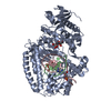
| Entry | Database: PDB / ID: 2hvh | |||||||||
|---|---|---|---|---|---|---|---|---|---|---|
| Title | ddCTP:O6MeG pair in the polymerase active site (0 position) | |||||||||
 Components Components |
| |||||||||
 Keywords Keywords | Transferase/DNA / DNA polymerase I / DNA replication / Klenow fragment / Protein-DNA complex / O6-methyl-guanine / Transferase-DNA COMPLEX | |||||||||
| Function / homology |  Function and homology information Function and homology information5'-3' exonuclease activity / 3'-5' exonuclease activity / DNA-templated DNA replication / double-strand break repair / DNA-directed DNA polymerase / DNA-directed DNA polymerase activity / nucleotide binding / DNA binding / metal ion binding Similarity search - Function | |||||||||
| Biological species |   Geobacillus stearothermophilus (bacteria) Geobacillus stearothermophilus (bacteria) | |||||||||
| Method |  X-RAY DIFFRACTION / SYNCHROTRON / FOURIER SYNTHESIS / Resolution: 2.492 Å X-RAY DIFFRACTION / SYNCHROTRON / FOURIER SYNTHESIS / Resolution: 2.492 Å | |||||||||
 Authors Authors | Warren, J.J. / Forsberg, L.J. / Beese, L.S. | |||||||||
 Citation Citation | Journal: Proc.Natl.Acad.Sci.Usa / Year: 2006 Title: The structural basis for the mutagenicity of O6-methyl-guanine lesions. Authors: Warren, J.J. / Forsberg, L.J. / Beese, L.S. | |||||||||
| History |
| |||||||||
| Remark 999 | SEQUENCE THE DNA POLYMERASE GENE WAS CLONED FROM AN ORGANISM THAT WAS CLASSIFIED AS A THERMOSTABLE ...SEQUENCE THE DNA POLYMERASE GENE WAS CLONED FROM AN ORGANISM THAT WAS CLASSIFIED AS A THERMOSTABLE STRAIN OF BACILLUS STEAROTHERMOPHILUS BY RIBOSOMAL RNA SEQUENCING. HOWEVER, THIS PARTICULAR GENE HAS MUCH GREATER HOMOLOGY WITH THE ANALOGOUS GENE FROM GEOBACILLUS KAUSTOPHILUS, UNP ACCESSION, Q5KWC1_GEOKA. |
- Structure visualization
| Structure viewer | Molecule: MolmilJmol/JSmol |
|---|
- Downloads & links
-Download
| PDBx/mmCIF format | 2hvh.cif.gz | 274.8 KB | Display | PDBx/mmCIF format |
|---|---|---|---|---|
| PDB format | pdb2hvh.ent.gz | 215.4 KB | Display | PDB format |
| PDBx/mmJSON format | 2hvh.json.gz | Tree view | PDBx/mmJSON format | |
| Others |  Other downloads Other downloads |
-Validation report
| Summary document | 2hvh_validation.pdf.gz | 2.1 MB | Display | wwPDB validaton report |
|---|---|---|---|---|
| Full document | 2hvh_full_validation.pdf.gz | 2.1 MB | Display | |
| Data in XML | 2hvh_validation.xml.gz | 46.3 KB | Display | |
| Data in CIF | 2hvh_validation.cif.gz | 65.4 KB | Display | |
| Arichive directory | https://data.pdbj.org/pub/pdb/validation_reports/hv/2hvhftp://data.pdbj.org/pub/pdb/validation_reports/hv/2hvh | HTTPS FTP |
-Related structure data
| Related structure data |  2hhqC  2hhsC 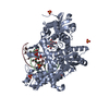 2hhtC  2hhuC  2hhvC  2hhwSC  2hhxC  2hviC  2hw3C  2hhy S: Starting model for refinement C: citing same article ( |
|---|---|
| Similar structure data |


-Links
 PDBj
PDBj


- Assembly
| Deposited unit | 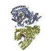
| ||||||||
|---|---|---|---|---|---|---|---|---|---|
| 1 | 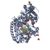
| ||||||||
| 2 | 
| ||||||||
| Unit cell |
| ||||||||
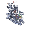
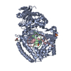
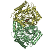
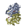
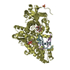
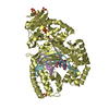
| Details | The asymmetric unit contains two complete biological assemblies, each of which consists of a DNA polymerase, two DNA strands, and an incoming ddCTP + Mn. |
-Components
-DNA chain , 2 types, 4 molecules BECF
| #1: DNA chain | Mass: 2675.775 Da / Num. of mol.: 2 / Source method: obtained synthetically / Details: DNA primer strand #2: DNA chain | Mass: 4030.650 Da / Num. of mol.: 2 / Source method: obtained synthetically / Details: DNA template strand |
|---|
-Protein / Sugars , 2 types, 5 molecules AD
| #3: Protein | Mass: 66202.945 Da / Num. of mol.: 2 Fragment: residues 299-876 (analogous to E. coli Klenow fragment) Source method: isolated from a genetically manipulated source Source: (gene. exp.) Geobacillus stearothermophilus (bacteria)Gene: polA / Plasmid: PUC / Species (production host): Escherichia coli / Production host: References: UniProt: Q5KWC1, UniProt: Q45458*PLUS, DNA-directed DNA polymerase #4: Polysaccharide |   Source method: isolated from a genetically manipulated source Details: oligosaccharide with reducing-end-to-reducing-end glycosidic bond References: sucrose |
|---|
-Non-polymers , 4 types, 287 molecules 

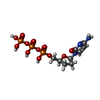

| #5: Chemical |  Mass: 54.938 Da / Num. of mol.: 2 / Source method: obtained synthetically / Formula: Mn Mass: 54.938 Da / Num. of mol.: 2 / Source method: obtained synthetically / Formula: Mn#6: Chemical | ChemComp-SO4 /  Mass: 96.063 Da / Num. of mol.: 5 / Source method: obtained synthetically / Formula: SO4 Mass: 96.063 Da / Num. of mol.: 5 / Source method: obtained synthetically / Formula: SO4#7: Chemical |  Type: DNA linking / Mass: 451.158 Da / Num. of mol.: 2 / Source method: obtained synthetically / Formula: C9H16N3O12P3 Type: DNA linking / Mass: 451.158 Da / Num. of mol.: 2 / Source method: obtained synthetically / Formula: C9H16N3O12P3#8: Water | ChemComp-HOH / | Mass: 18.015 Da / Num. of mol.: 278 / Source method: isolated from a natural source / Formula: H2O |
|---|
-Experimental details
-Experiment
| Experiment | Method: X-RAY DIFFRACTION / Number of used crystals: 1 |
|---|
- Sample preparation
| Crystal | Density Matthews: 2.66 Å3/Da / Density % sol: 53.78 % | ||||||||||||||||||||||||||||||||||||||||||||
|---|---|---|---|---|---|---|---|---|---|---|---|---|---|---|---|---|---|---|---|---|---|---|---|---|---|---|---|---|---|---|---|---|---|---|---|---|---|---|---|---|---|---|---|---|---|
| Crystal grow | Temperature: 290 K / Method: vapor diffusion, hanging drop / pH: 5.8 Details: 55% saturated ammonium sulfate, 100 mM MES, 2.5% MPD, 10mM MnSO4, 10mM MgSO4, pH 5.8, VAPOR DIFFUSION, HANGING DROP, temperature 290K | ||||||||||||||||||||||||||||||||||||||||||||
| Components of the solutions |
|
-Data collection
| Diffraction | Mean temperature: 100 K |
|---|---|
| Diffraction source | Source: SYNCHROTRON / Site: APS  / Beamline: 22-ID / Wavelength: 1 Å / Beamline: 22-ID / Wavelength: 1 Å |
| Detector | Type: MARMOSAIC 300 mm CCD / Detector: CCD / Date: Jul 7, 2006 / Details: mirrors |
| Radiation | Monochromator: Si 111 / Protocol: SINGLE WAVELENGTH / Monochromatic (M) / Laue (L): M / Scattering type: x-ray |
| Radiation wavelength | Wavelength: 1 Å / Relative weight: 1 |
| Reflection | Resolution: 2.492→47.298 Å / Num. all: 54569 / Num. obs: 54569 / % possible obs: 98.8 % / Observed criterion σ(I): -3 / Redundancy: 5.6 % / Rmerge(I) obs: 0.135 / Rsym value: 0.135 / Net I/σ(I): 11.4 |
| Reflection shell | Resolution: 2.492→2.63 Å / Redundancy: 4.9 % / Rmerge(I) obs: 0.414 / Mean I/σ(I) obs: 3.2 / Num. measured all: 36059 / Num. unique all: 7420 / Rsym value: 0.414 / % possible all: 93.3 |
- Processing
| Software |
| ||||||||||||||||||||||||||||||||||||||||||||||||||||||||||||||||||||||||||||||||||||||||||
|---|---|---|---|---|---|---|---|---|---|---|---|---|---|---|---|---|---|---|---|---|---|---|---|---|---|---|---|---|---|---|---|---|---|---|---|---|---|---|---|---|---|---|---|---|---|---|---|---|---|---|---|---|---|---|---|---|---|---|---|---|---|---|---|---|---|---|---|---|---|---|---|---|---|---|---|---|---|---|---|---|---|---|---|---|---|---|---|---|---|---|---|
| Refinement | Method to determine structure: FOURIER SYNTHESIS Starting model: pdb entry 2HHW Resolution: 2.492→47.298 Å / Cor.coef. Fo:Fc: 0.924 / Cor.coef. Fo:Fc free: 0.883 / SU B: 10.804 / SU ML: 0.236 / Isotropic thermal model: Isotropic / Cross valid method: THROUGHOUT / σ(F): 0 / ESU R: 0.537 / ESU R Free: 0.301 / Stereochemistry target values: MAXIMUM LIKELIHOOD / Details: HYDROGENS HAVE BEEN ADDED IN THE RIDING POSITIONS
| ||||||||||||||||||||||||||||||||||||||||||||||||||||||||||||||||||||||||||||||||||||||||||
| Solvent computation | Ion probe radii: 0.8 Å / Shrinkage radii: 0.8 Å / VDW probe radii: 1.2 Å / Solvent model: BABINET MODEL WITH MASK | ||||||||||||||||||||||||||||||||||||||||||||||||||||||||||||||||||||||||||||||||||||||||||
| Displacement parameters | Biso mean: 31.483 Å2
| ||||||||||||||||||||||||||||||||||||||||||||||||||||||||||||||||||||||||||||||||||||||||||
| Refinement step | Cycle: LAST / Resolution: 2.492→47.298 Å
| ||||||||||||||||||||||||||||||||||||||||||||||||||||||||||||||||||||||||||||||||||||||||||
| Refine LS restraints |
| ||||||||||||||||||||||||||||||||||||||||||||||||||||||||||||||||||||||||||||||||||||||||||
| LS refinement shell | Resolution: 2.492→2.557 Å / Total num. of bins used: 20
|
