 Movie
Movie Controller


+ Open data

- Basic information
| Entry | Database: PDB / ID: 2hr7 | |||||||||
|---|---|---|---|---|---|---|---|---|---|---|
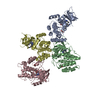
| Title | Insulin receptor (domains 1-3) | |||||||||
 Components Components | Insulin receptor | |||||||||
 Keywords Keywords | TRANSFERASE / HORMONE RECEPTOR / LEUCINE RICH REPEAT | |||||||||
| Function / homology |  Function and homology information Function and homology informationregulation of female gonad development / positive regulation of meiotic cell cycle / insulin-like growth factor II binding / positive regulation of developmental growth / male sex determination / insulin receptor complex / insulin-like growth factor I binding / insulin receptor activity / positive regulation of protein-containing complex disassembly / exocrine pancreas development ...regulation of female gonad development / positive regulation of meiotic cell cycle / insulin-like growth factor II binding / positive regulation of developmental growth / male sex determination / insulin receptor complex / insulin-like growth factor I binding / insulin receptor activity / positive regulation of protein-containing complex disassembly / exocrine pancreas development / dendritic spine maintenance / adrenal gland development / insulin binding / cargo receptor activity / PTB domain binding / Signaling by Insulin receptor / IRS activation / neuronal cell body membrane / positive regulation of respiratory burst / amyloid-beta clearance / regulation of embryonic development / insulin receptor substrate binding / positive regulation of receptor internalization / epidermis development / positive regulation of glycogen biosynthetic process / Signal attenuation / protein kinase activator activity / transport across blood-brain barrier / phosphatidylinositol 3-kinase binding / heart morphogenesis / Insulin receptor recycling / insulin-like growth factor receptor binding / neuron projection maintenance / positive regulation of mitotic nuclear division / receptor-mediated endocytosis / Insulin receptor signalling cascade / dendrite membrane / positive regulation of glycolytic process / positive regulation of D-glucose import across plasma membrane / learning / receptor protein-tyrosine kinase / caveola / receptor internalization / cellular response to growth factor stimulus / male gonad development / cellular response to insulin stimulus / memory / positive regulation of nitric oxide biosynthetic process / insulin receptor signaling pathway / protein autophosphorylation / late endosome / glucose homeostasis / amyloid-beta binding / PI5P, PP2A and IER3 Regulate PI3K/AKT Signaling / protein tyrosine kinase activity / positive regulation of MAPK cascade / positive regulation of canonical NF-kappaB signal transduction / positive regulation of phosphatidylinositol 3-kinase/protein kinase B signal transduction / lysosome / signaling receptor complex / endosome membrane / positive regulation of cell migration / G protein-coupled receptor signaling pathway / external side of plasma membrane / protein domain specific binding / axon / positive regulation of cell population proliferation / symbiont entry into host cell / regulation of DNA-templated transcription / positive regulation of DNA-templated transcription / GTP binding / protein-containing complex binding / extracellular exosome / ATP binding / membrane / identical protein binding / plasma membrane Similarity search - Function | |||||||||
| Biological species |  Homo sapiens (human) Homo sapiens (human) | |||||||||
| Method |  X-RAY DIFFRACTION / SYNCHROTRON / MOLECULAR REPLACEMENT / Resolution: 2.32 Å X-RAY DIFFRACTION / SYNCHROTRON / MOLECULAR REPLACEMENT / Resolution: 2.32 Å | |||||||||
 Authors Authors | Garrett, T.P.J. / Ward, C.W. | |||||||||
 Citation Citation | Journal: Proc.Natl.Acad.Sci.Usa / Year: 2006 Title: The first three domains of the insulin receptor differ structurally from the insulin-like growth factor 1 receptor in the regions governing ligand specificity. Authors: Lou, M. / Garrett, T.P. / McKern, N.M. / Hoyne, P.A. / Epa, V.C. / Bentley, J.D. / Lovrecz, G.O. / Cosgrove, L.J. / Frenkel, M.J. / Ward, C.W. | |||||||||
| History |
| |||||||||
| Remark 999 | SEQUENCE The protein was treated with AspN protease and the most probable C-terminal residue is 486. |
- Structure visualization
| Structure viewer | Molecule: MolmilJmol/JSmol |
|---|
- Downloads & links
-Download
| PDBx/mmCIF format | 2hr7.cif.gz | 227.4 KB | Display | PDBx/mmCIF format |
|---|---|---|---|---|
| PDB format | pdb2hr7.ent.gz | 184.5 KB | Display | PDB format |
| PDBx/mmJSON format | 2hr7.json.gz | Tree view | PDBx/mmJSON format | |
| Others |  Other downloads Other downloads |
-Validation report
| Arichive directory | https://data.pdbj.org/pub/pdb/validation_reports/hr/2hr7ftp://data.pdbj.org/pub/pdb/validation_reports/hr/2hr7 | HTTPS FTP |
|---|
-Related structure data
| Related structure data | 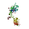 1igrS S: Starting model for refinement |
|---|---|
| Similar structure data |


-Links
 PDBj
PDBj


- Assembly
| Deposited unit | 
| ||||||||
|---|---|---|---|---|---|---|---|---|---|
| 1 |
| ||||||||
| Unit cell |
| ||||||||
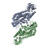
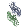
| Details | The biological assembly is a disulfide-liked dimer where the linking residues are not in this fragment. The two molecules in the asymmetric unit are not likely to be in the correct orientation for the dimer. |
-Components
-Protein , 1 types, 2 molecules AB
| #1: Protein | Mass: 55544.707 Da / Num. of mol.: 2 / Fragment: Domains 1-3 (residues 28-512) Source method: isolated from a genetically manipulated source Source: (gene. exp.) Homo sapiens (human) / Gene: INSR / Plasmid: PEE14-IR485 / Production host:   Cricetulus griseus (Chinese hamster) / Strain (production host): Lec8 / References: UniProt: P06213, EC: 2.7.1.112 Cricetulus griseus (Chinese hamster) / Strain (production host): Lec8 / References: UniProt: P06213, EC: 2.7.1.112 |
|---|
-Sugars , 7 types, 15 molecules 
| #2: Polysaccharide | 2-acetamido-2-deoxy-beta-D-glucopyranose-(1-4)-2-acetamido-2-deoxy-beta-D-glucopyranose Source method: isolated from a genetically manipulated source | ||||||||||
|---|---|---|---|---|---|---|---|---|---|---|---|
| #3: Polysaccharide | Source method: isolated from a genetically manipulated source #4: Polysaccharide | beta-D-mannopyranose-(1-4)-2-acetamido-2-deoxy-beta-D-glucopyranose-(1-4)-2-acetamido-2-deoxy-beta- ...beta-D-mannopyranose-(1-4)-2-acetamido-2-deoxy-beta-D-glucopyranose-(1-4)-2-acetamido-2-deoxy-beta-D-glucopyranose | Source method: isolated from a genetically manipulated source #5: Polysaccharide | Source method: isolated from a genetically manipulated source #6: Polysaccharide | 2-acetamido-2-deoxy-beta-D-glucopyranose-(1-4)-[alpha-L-fucopyranose-(1-6)]2-acetamido-2-deoxy-beta- ...2-acetamido-2-deoxy-beta-D-glucopyranose-(1-4)-[alpha-L-fucopyranose-(1-6)]2-acetamido-2-deoxy-beta-D-glucopyranose Source method: isolated from a genetically manipulated source #7: Polysaccharide | alpha-D-mannopyranose-(1-3)-[alpha-D-mannopyranose-(1-6)]alpha-D-mannopyranose-(1-6)-[alpha-D- ...alpha-D-mannopyranose-(1-3)-[alpha-D-mannopyranose-(1-6)]alpha-D-mannopyranose-(1-6)-[alpha-D-mannopyranose-(1-3)]beta-D-mannopyranose-(1-4)-2-acetamido-2-deoxy-beta-D-glucopyranose-(1-4)-2-acetamido-2-deoxy-beta-D-glucopyranose | Source method: isolated from a genetically manipulated source #8: Sugar | ChemComp-NAG /  Type: D-saccharide, beta linking / Mass: 221.208 Da / Num. of mol.: 4 Type: D-saccharide, beta linking / Mass: 221.208 Da / Num. of mol.: 4Source method: isolated from a genetically manipulated source Formula: C8H15NO6 |
-Non-polymers , 4 types, 399 molecules 


| #9: Chemical | ChemComp-SO4 /  Mass: 96.063 Da / Num. of mol.: 8 / Source method: obtained synthetically / Formula: SO4 Mass: 96.063 Da / Num. of mol.: 8 / Source method: obtained synthetically / Formula: SO4#10: Chemical | ChemComp-P33 / |  Mass: 326.383 Da / Num. of mol.: 1 / Source method: obtained synthetically / Formula: C14H30O8 / Comment: precipitant*YM Mass: 326.383 Da / Num. of mol.: 1 / Source method: obtained synthetically / Formula: C14H30O8 / Comment: precipitant*YM#11: Chemical | ChemComp-GOL /  Mass: 92.094 Da / Num. of mol.: 19 / Source method: obtained synthetically / Formula: C3H8O3 Mass: 92.094 Da / Num. of mol.: 19 / Source method: obtained synthetically / Formula: C3H8O3#12: Water | ChemComp-HOH / | Mass: 18.015 Da / Num. of mol.: 371 / Source method: isolated from a natural source / Formula: H2O |
|---|
-Details
| Has protein modification | Y |
|---|
-Experimental details
-Experiment
| Experiment | Method: X-RAY DIFFRACTION / Number of used crystals: 1 |
|---|
- Sample preparation
| Crystal | Density Matthews: 4.74 Å3/Da / Density % sol: 74.03 % |
|---|---|
| Crystal grow | Temperature: 291 K / pH: 8.5 Details: 1.5-1.65 M ammonium sulfate, 2 % PEG 400, pH 8.5, VAPOR DIFFUSION, HANGING DROP, temperature 291K, pH 8.50 |
-Data collection
| Diffraction | Mean temperature: 103 K |
|---|---|
| Diffraction source | Source: SYNCHROTRON / Site: APS  / Beamline: 14-BM-D / Wavelength: 1.037 / Beamline: 14-BM-D / Wavelength: 1.037 |
| Detector | Type: MAR scanner 345 mm plate / Detector: IMAGE PLATE / Date: Jul 12, 1998 |
| Radiation | Monochromator: SI(111) DOUBLE-CRYSTAL / Protocol: SINGLE WAVELENGTH / Monochromatic (M) / Laue (L): M / Scattering type: x-ray |
| Radiation wavelength | Wavelength: 1.037 Å / Relative weight: 1 |
| Reflection | Resolution: 2.3→99 Å / Num. obs: 94059 / % possible obs: 99.6 % / Observed criterion σ(I): -5 / Redundancy: 3.4 % / Rmerge(I) obs: 0.072 / Net I/σ(I): 12 |
| Reflection shell | Resolution: 2.3→2.38 Å / Redundancy: 3.2 % / Rmerge(I) obs: 0.77 / Mean I/σ(I) obs: 2.1 / % possible all: 99.3 |
- Processing
| Software |
| ||||||||||||||||||||||||||||||||||||||||||||||||||||||||||||||||||||||||||||||||||||||||||||||||||||||||||||||||||||||||||||||||||||||||||||||||||||||||||||||||||||||||||
|---|---|---|---|---|---|---|---|---|---|---|---|---|---|---|---|---|---|---|---|---|---|---|---|---|---|---|---|---|---|---|---|---|---|---|---|---|---|---|---|---|---|---|---|---|---|---|---|---|---|---|---|---|---|---|---|---|---|---|---|---|---|---|---|---|---|---|---|---|---|---|---|---|---|---|---|---|---|---|---|---|---|---|---|---|---|---|---|---|---|---|---|---|---|---|---|---|---|---|---|---|---|---|---|---|---|---|---|---|---|---|---|---|---|---|---|---|---|---|---|---|---|---|---|---|---|---|---|---|---|---|---|---|---|---|---|---|---|---|---|---|---|---|---|---|---|---|---|---|---|---|---|---|---|---|---|---|---|---|---|---|---|---|---|---|---|---|---|---|---|---|---|
| Refinement | Method to determine structure: MOLECULAR REPLACEMENT Starting model: PDB ENTRY 1IGR Resolution: 2.32→40 Å / Cor.coef. Fo:Fc: 0.951 / Cor.coef. Fo:Fc free: 0.932 / SU B: 5.035 / SU ML: 0.122 / Cross valid method: THROUGHOUT / σ(F): 0 / ESU R: 0.183 / ESU R Free: 0.17 / Stereochemistry target values: ENGH & HUBER / Details: HYDROGENS HAVE BEEN ADDED IN THE RIDING POSITIONS
| ||||||||||||||||||||||||||||||||||||||||||||||||||||||||||||||||||||||||||||||||||||||||||||||||||||||||||||||||||||||||||||||||||||||||||||||||||||||||||||||||||||||||||
| Solvent computation | Ion probe radii: 0.8 Å / Shrinkage radii: 0.8 Å / VDW probe radii: 1.2 Å / Solvent model: BABINET MODEL WITH MASK | ||||||||||||||||||||||||||||||||||||||||||||||||||||||||||||||||||||||||||||||||||||||||||||||||||||||||||||||||||||||||||||||||||||||||||||||||||||||||||||||||||||||||||
| Displacement parameters | Biso mean: 47.35 Å2
| ||||||||||||||||||||||||||||||||||||||||||||||||||||||||||||||||||||||||||||||||||||||||||||||||||||||||||||||||||||||||||||||||||||||||||||||||||||||||||||||||||||||||||
| Refinement step | Cycle: LAST / Resolution: 2.32→40 Å
| ||||||||||||||||||||||||||||||||||||||||||||||||||||||||||||||||||||||||||||||||||||||||||||||||||||||||||||||||||||||||||||||||||||||||||||||||||||||||||||||||||||||||||
| Refine LS restraints |
| ||||||||||||||||||||||||||||||||||||||||||||||||||||||||||||||||||||||||||||||||||||||||||||||||||||||||||||||||||||||||||||||||||||||||||||||||||||||||||||||||||||||||||
| LS refinement shell | Resolution: 2.32→2.38 Å / Total num. of bins used: 20
|
