[English] 日本語
Yorodumi
- PDB-2h9e: Crystal Structure of FXa/selectide/NAPC2 ternary complex -

+
Open data


ID or keywords:

Loading...

-
Basic information

Entry
Database: PDB / ID: 2h9e
TitleCrystal Structure of FXa/selectide/NAPC2 ternary complex
Components
  • (Coagulation factor X ...) x 2
  • Anti-coagulant protein C2
  • selectide inhibitor DTY-ILE-ARG-LEU-LPD peptide
KeywordsHYDROLASE/HYDROLASE INHIBITOR / factor Xa / NAPc2 / selectide / hydrolase-hydrolase inhibitor complex / blood clotting
Function / homology
Function and homology information


coagulation factor Xa / Defective factor IX causes thrombophilia / Defective cofactor function of FVIIIa variant / Defective F9 variant does not activate FX / Extrinsic Pathway of Fibrin Clot Formation / positive regulation of leukocyte chemotaxis / positive regulation of TOR signaling / Gamma-carboxylation of protein precursors / Transport of gamma-carboxylated protein precursors from the endoplasmic reticulum to the Golgi apparatus / Common Pathway of Fibrin Clot Formation ...coagulation factor Xa / Defective factor IX causes thrombophilia / Defective cofactor function of FVIIIa variant / Defective F9 variant does not activate FX / Extrinsic Pathway of Fibrin Clot Formation / positive regulation of leukocyte chemotaxis / positive regulation of TOR signaling / Gamma-carboxylation of protein precursors / Transport of gamma-carboxylated protein precursors from the endoplasmic reticulum to the Golgi apparatus / Common Pathway of Fibrin Clot Formation / Removal of aminoterminal propeptides from gamma-carboxylated proteins / Intrinsic Pathway of Fibrin Clot Formation / serine-type endopeptidase inhibitor activity / phospholipid binding / Golgi lumen / blood coagulation / positive regulation of cell migration / endoplasmic reticulum lumen / external side of plasma membrane / serine-type endopeptidase activity / calcium ion binding / proteolysis / extracellular space / extracellular region / plasma membrane
Similarity search - Function
Trypsin Inhibitor-like, cysteine rich domain / Serine protease inhibitor-like superfamily / Trypsin Inhibitor like cysteine rich domain / Peptidase S1A, coagulation factor VII/IX/X/C/Z / Coagulation factor-like, Gla domain superfamily / Laminin / Coagulation Factor Xa inhibitory site / Laminin / EGF-like domain / EGF-type aspartate/asparagine hydroxylation site ...Trypsin Inhibitor-like, cysteine rich domain / Serine protease inhibitor-like superfamily / Trypsin Inhibitor like cysteine rich domain / Peptidase S1A, coagulation factor VII/IX/X/C/Z / Coagulation factor-like, Gla domain superfamily / Laminin / Coagulation Factor Xa inhibitory site / Laminin / EGF-like domain / EGF-type aspartate/asparagine hydroxylation site / EGF-like calcium-binding, conserved site / Calcium-binding EGF-like domain signature. / Aspartic acid and asparagine hydroxylation site. / EGF-like calcium-binding domain / Calcium-binding EGF-like domain / Vitamin K-dependent carboxylation/gamma-carboxyglutamic (GLA) domain / Gamma-carboxyglutamic acid-rich (GLA) domain / Gamma-carboxyglutamic acid-rich (GLA) domain superfamily / Vitamin K-dependent carboxylation domain. / Gla domain profile. / Domain containing Gla (gamma-carboxyglutamate) residues. / Epidermal growth factor-like domain. / EGF-like domain profile. / EGF-like domain signature 2. / EGF-like domain signature 1. / EGF-like domain / Ribbon / Serine proteases, trypsin family, histidine active site / Serine proteases, trypsin family, serine active site / Peptidase S1A, chymotrypsin family / Serine proteases, trypsin family, histidine active site. / Serine proteases, trypsin domain profile. / Serine proteases, trypsin family, serine active site. / Trypsin-like serine protease / Serine proteases, trypsin domain / Trypsin / Trypsin-like serine proteases / Thrombin, subunit H / Peptidase S1, PA clan, chymotrypsin-like fold / Peptidase S1, PA clan / Beta Barrel / Mainly Beta
Similarity search - Domain/homology
ACETATE ION / PHOSPHATE ION / Coagulation factor X / Anti-coagulant protein C2
Similarity search - Component
Biological speciesAncylostoma caninum (dog hookworm)
Homo sapiens (human)
MethodX-RAY DIFFRACTION / SYNCHROTRON / MOLECULAR REPLACEMENT / Resolution: 2.2 Å
AuthorsMurakami, M.T. / Geiger, G. / Tulinsky, A. / Arni, R.K.
CitationJournal: J.Mol.Biol. / Year: 2007
Title: Intermolecular Interactions and Characterization of the Novel Factor Xa Exosite Involved in Macromolecular Recognition and Inhibition: Crystal Structure of Human Gla-domainless Factor Xa ...Title: Intermolecular Interactions and Characterization of the Novel Factor Xa Exosite Involved in Macromolecular Recognition and Inhibition: Crystal Structure of Human Gla-domainless Factor Xa Complexed with the Anticoagulant Protein NAPc2 from the Hematophagous Nematode Ancylostoma caninum.
Authors: Murakami, M.T. / Rios-Steiner, J. / Weaver, S.E. / Tulinsky, A. / Geiger, J.H. / Arni, R.K.
History
DepositionJun 9, 2006Deposition site: RCSB / Processing site: RCSB
Revision 1.0Feb 13, 2007Provider: repository / Type: Initial release
Revision 1.1May 1, 2008Group: Version format compliance
Revision 1.2Jul 13, 2011Group: Atomic model / Database references ...Atomic model / Database references / Derived calculations / Non-polymer description / Structure summary / Version format compliance
Revision 1.3Oct 18, 2017Group: Refinement description / Category: software / Item: _software.name

-
Structure visualization

Structure viewerMolecule:
MolmilJmol/JSmol

Downloads & links

-
Assembly

Deposited unit
H: Coagulation factor X heavy chain
L: Coagulation factor X light chain
C: Anti-coagulant protein C2
S: selectide inhibitor DTY-ILE-ARG-LEU-LPD peptide
hetero molecules


Theoretical massNumber of molelcules
Total (without water)53,80414
Polymers53,0344
Non-polymers77010
Water4,648258
1


  • Idetical with deposited unit
  • defined by author&software
TypeNameSymmetry operationNumber
identity operation1_555x,y,z1
Buried area6490 Å2
ΔGint-60 kcal/mol
Surface area15610 Å2
MethodPISA
Unit cell
Length a, b, c (Å)48.945, 86.410, 145.892
Angle α, β, γ (deg.)90.00, 90.00, 90.00
Int Tables number19
Space group name H-MP212121
Details
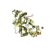
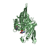
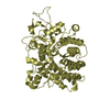
The biological assembly is the chain H (catalytic domain or heavy chain) and chain L (EGF2 domain or light chain), which forms the Gla-domainless factor Xa

-
Components

-
Coagulation factor X ... , 2 types, 2 molecules HL

#1: Protein Coagulation factor X heavy chain


Mass: 26346.000 Da / Num. of mol.: 1 / Fragment: catalytic domain / Source method: isolated from a natural source / Source: (natural) Homo sapiens (human) / References: UniProt: P00742, coagulation factor Xa
#2: Protein Coagulation factor X light chain


Mass: 16366.871 Da / Num. of mol.: 1 / Fragment: EGF-like 1 domain / Source method: isolated from a natural source / Source: (natural) Homo sapiens (human) / References: UniProt: P00742, coagulation factor Xa

-
Protein / Protein/peptide , 2 types, 2 molecules CS

#3: Protein Anti-coagulant protein C2


Mass: 9660.605 Da / Num. of mol.: 1
Source method: isolated from a genetically manipulated source
Source: (gene. exp.) Ancylostoma caninum (dog hookworm) / Production host: Escherichia coli (E. coli) / References: UniProt: Q16938
#4: Protein/peptide selectide inhibitor DTY-ILE-ARG-LEU-LPD peptide


Mass: 660.829 Da / Num. of mol.: 1 / Source method: obtained synthetically / Details: THE PEPTIDE WAS CHEMICALLY SYNTHESIZED.

-
Non-polymers , 4 types, 268 molecules

#5: Chemical
ChemComp-PO4 / PHOSPHATE ION


Mass: 94.971 Da / Num. of mol.: 6 / Source method: obtained synthetically / Formula: PO4
#6: Chemical ChemComp-ACT / ACETATE ION


Mass: 59.044 Da / Num. of mol.: 3 / Source method: obtained synthetically / Formula: C2H3O2
#7: Chemical ChemComp-NA / SODIUM ION


Mass: 22.990 Da / Num. of mol.: 1 / Source method: obtained synthetically / Formula: Na
#8: Water ChemComp-HOH / water


Mass: 18.015 Da / Num. of mol.: 258 / Source method: isolated from a natural source / Formula: H2O

-
Experimental details

-
Experiment

ExperimentMethod: X-RAY DIFFRACTION / Number of used crystals: 2

-
Sample preparation

CrystalDensity Matthews: 2.9 Å3/Da / Density % sol: 57.54 %
Crystal growTemperature: 293 K / Method: vapor diffusion, hanging drop / pH: 5.6
Details: peg 8000, potassium dihydrogen phosphate, acetate ion, pH 5.6, VAPOR DIFFUSION, HANGING DROP, temperature 293K

-
Data collection

DiffractionMean temperature: 100 K
Diffraction sourceSource: SYNCHROTRON / Site: APS / Beamline: 14-BM-C / Wavelength: 1.01 Å
DetectorType: MAR CCD 165 mm / Detector: CCD / Date: Jul 9, 2001
RadiationProtocol: SINGLE WAVELENGTH / Monochromatic (M) / Laue (L): M / Scattering type: x-ray
Radiation wavelengthWavelength: 1.01 Å / Relative weight: 1
ReflectionResolution: 2.2→37.17 Å / Num. all: 32280 / Num. obs: 32172 / % possible obs: 99.6 % / Observed criterion σ(F): 1 / Observed criterion σ(I): 2
Reflection shellResolution: 2.2→2.26 Å / % possible all: 99.5

-
Processing

Software
NameVersionClassification
REFMAC5.2.0005refinement
MAR345data collection
AMoREphasing
RefinementMethod to determine structure: MOLECULAR REPLACEMENT / Resolution: 2.2→74.33 Å / Cor.coef. Fo:Fc: 0.939 / Cor.coef. Fo:Fc free: 0.915 / SU B: 4.984 / SU ML: 0.131 / Cross valid method: THROUGHOUT / σ(F): 1 / ESU R: 0.195 / ESU R Free: 0.187 / Stereochemistry target values: MAXIMUM LIKELIHOOD
RfactorNum. reflection% reflectionSelection details
Rfree0.26832 1631 5.1 %RANDOM
Rwork0.21959 ---
all0.233 32172 --
obs0.22209 30145 99.48 %-
Solvent computationIon probe radii: 0.8 Å / Shrinkage radii: 0.8 Å / VDW probe radii: 1.2 Å / Solvent model: MASK
Displacement parametersBiso mean: 45.472 Å2
Baniso -1Baniso -2Baniso -3
1--4.36 Å20 Å20 Å2
2---0.95 Å20 Å2
3---5.31 Å2
Refinement stepCycle: LAST / Resolution: 2.2→74.33 Å
ProteinNucleic acidLigandSolventTotal
Num. atoms2637 0 43 258 2938
Refine LS restraints
Refine-IDTypeDev idealDev ideal targetNumber
X-RAY DIFFRACTIONr_bond_refined_d0.0240.0222736
X-RAY DIFFRACTIONr_angle_refined_deg2.1421.9613693
X-RAY DIFFRACTIONr_dihedral_angle_1_deg8.695332
X-RAY DIFFRACTIONr_dihedral_angle_2_deg37.97624.436133
X-RAY DIFFRACTIONr_dihedral_angle_3_deg21.02915458
X-RAY DIFFRACTIONr_dihedral_angle_4_deg18.9331517
X-RAY DIFFRACTIONr_chiral_restr0.2160.2385
X-RAY DIFFRACTIONr_gen_planes_refined0.010.022078
X-RAY DIFFRACTIONr_nbd_refined0.2440.21280
X-RAY DIFFRACTIONr_nbtor_refined0.3190.21768
X-RAY DIFFRACTIONr_xyhbond_nbd_refined0.2540.2253
X-RAY DIFFRACTIONr_symmetry_vdw_refined0.2660.221
X-RAY DIFFRACTIONr_symmetry_hbond_refined0.2430.25
X-RAY DIFFRACTIONr_mcbond_it1.3391.51676
X-RAY DIFFRACTIONr_mcangle_it2.35422676
X-RAY DIFFRACTIONr_scbond_it3.40331094
X-RAY DIFFRACTIONr_scangle_it4.954.51017
LS refinement shellResolution: 2.2→2.255 Å / Total num. of bins used: 20
RfactorNum. reflection% reflection
Rfree0.339 127 -
Rwork0.241 2186 -
obs--98.47 %

+
About Yorodumi

-
News

-
Feb 9, 2022. New format data for meta-information of EMDB entries

New format data for meta-information of EMDB entries

  • Version 3 of the EMDB header file is now the official format.
  • The previous official version 1.9 will be removed from the archive.

Related info.:EMDB header

External links:wwPDB to switch to version 3 of the EMDB data model

-
Aug 12, 2020. Covid-19 info

Covid-19 info

URL: https://pdbjlvh1.pdbj.org/emnavi/covid19.php

New page: Covid-19 featured information page in EM Navigator.

Related info.:Covid-19 info / Mar 5, 2020. Novel coronavirus structure data

+
Mar 5, 2020. Novel coronavirus structure data

Novel coronavirus structure data

Related info.:Yorodumi Speices / Aug 12, 2020. Covid-19 info

External links:COVID-19 featured content - PDBj / Molecule of the Month (242):Coronavirus Proteases

+
Jan 31, 2019. EMDB accession codes are about to change! (news from PDBe EMDB page)

EMDB accession codes are about to change! (news from PDBe EMDB page)

  • The allocation of 4 digits for EMDB accession codes will soon come to an end. Whilst these codes will remain in use, new EMDB accession codes will include an additional digit and will expand incrementally as the available range of codes is exhausted. The current 4-digit format prefixed with “EMD-” (i.e. EMD-XXXX) will advance to a 5-digit format (i.e. EMD-XXXXX), and so on. It is currently estimated that the 4-digit codes will be depleted around Spring 2019, at which point the 5-digit format will come into force.
  • The EM Navigator/Yorodumi systems omit the EMD- prefix.

Related info.:Q: What is EMD? / ID/Accession-code notation in Yorodumi/EM Navigator

External links:EMDB Accession Codes are Changing Soon! / Contact to PDBj

+
Jul 12, 2017. Major update of PDB

Major update of PDB

  • wwPDB released updated PDB data conforming to the new PDBx/mmCIF dictionary.
  • This is a major update changing the version number from 4 to 5, and with Remediation, in which all the entries are updated.
  • In this update, many items about electron microscopy experimental information are reorganized (e.g. em_software).
  • Now, EM Navigator and Yorodumi are based on the updated data.

External links:wwPDB Remediation / Enriched Model Files Conforming to OneDep Data Standards Now Available in the PDB FTP Archive

-
Yorodumi

Thousand views of thousand structures

  • Yorodumi is a browser for structure data from EMDB, PDB, SASBDB, etc.
  • This page is also the successor to EM Navigator detail page, and also detail information page/front-end page for Omokage search.
  • The word "yorodu" (or yorozu) is an old Japanese word meaning "ten thousand". "mi" (miru) is to see.

Related info.:EMDB / PDB / SASBDB / Comparison of 3 databanks / Yorodumi Search / Aug 31, 2016. New EM Navigator & Yorodumi / Yorodumi Papers / Jmol/JSmol / Function and homology information / Changes in new EM Navigator and Yorodumi

Read more