[English] 日本語
Yorodumi
- PDB-2h8a: Structure of Microsomal Glutathione Transferase 1 in Complex with... -

+
Open data


ID or keywords:

Loading...

-
Basic information

Entry
Database: PDB / ID: 2h8a
TitleStructure of Microsomal Glutathione Transferase 1 in Complex with Glutathione
ComponentsMicrosomal glutathione S-transferase 1
KeywordsTRANSFERASE / Membrane protein
Function / homology
Function and homology information


cellular response to lipid hydroperoxide / Aflatoxin activation and detoxification / glutathione transport / Glutathione conjugation / glutathione binding / Leydig cell differentiation / glutathione peroxidase activity / peroxisomal membrane / Neutrophil degranulation / glutathione transferase ...cellular response to lipid hydroperoxide / Aflatoxin activation and detoxification / glutathione transport / Glutathione conjugation / glutathione binding / Leydig cell differentiation / glutathione peroxidase activity / peroxisomal membrane / Neutrophil degranulation / glutathione transferase / glutathione transferase activity / : / glutathione metabolic process / apical part of cell / mitochondrial outer membrane / response to lipopolysaccharide / response to xenobiotic stimulus / endoplasmic reticulum membrane / endoplasmic reticulum / mitochondrion / identical protein binding / membrane
Similarity search - Function
Microsomal glutathione S-transferase 1-like / Membrane associated eicosanoid/glutathione metabolism-like domain / Membrane-associated, eicosanoid/glutathione metabolism (MAPEG) protein / Membrane associated eicosanoid/glutathione metabolism-like domain superfamily / MAPEG family / Four Helix Bundle (Hemerythrin (Met), subunit A) / Up-down Bundle / Mainly Alpha
Similarity search - Domain/homology
GLUTATHIONE / Microsomal glutathione S-transferase 1
Similarity search - Component
Biological speciesRattus norvegicus (Norway rat)
MethodELECTRON CRYSTALLOGRAPHY / electron crystallography / cryo EM / Resolution: 3.2 Å
AuthorsHebert, H.
Citation
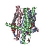
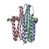
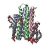
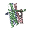
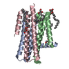
Journal: J Mol Biol / Year: 2006
Title: Structural basis for detoxification and oxidative stress protection in membranes.
Authors: Peter J Holm / Priyaranjan Bhakat / Caroline Jegerschöld / Nobuhiko Gyobu / Kaoru Mitsuoka / Yoshinori Fujiyoshi / Ralf Morgenstern / Hans Hebert /
Abstract: Synthesis of mediators of fever, pain and inflammation as well as protection against reactive molecules and oxidative stress is a hallmark of the MAPEG superfamily (membrane associated proteins in ...Synthesis of mediators of fever, pain and inflammation as well as protection against reactive molecules and oxidative stress is a hallmark of the MAPEG superfamily (membrane associated proteins in eicosanoid and glutathione metabolism). The structure of a MAPEG member, rat microsomal glutathione transferase 1, at 3.2 A resolution, solved here in complex with glutathione by electron crystallography, defines the active site location and a cytosolic domain involved in enzyme activation. The glutathione binding site is found to be different from that of the canonical soluble glutathione transferases. The architecture of the homotrimer supports a catalytic mechanism involving subunit interactions and reveals both cytosolic and membraneous substrate entry sites, providing a rationale for the membrane location of the enzyme.
History
DepositionJun 7, 2006Deposition site: RCSB / Processing site: RCSB
Revision 1.0May 22, 2007Provider: repository / Type: Initial release
Revision 1.1May 1, 2008Group: Version format compliance
Revision 1.2Jul 13, 2011Group: Derived calculations / Version format compliance
Revision 1.3Jan 18, 2012Group: Non-polymer description
Revision 1.4Mar 21, 2012Group: Non-polymer description
Revision 1.5Feb 14, 2024Group: Author supporting evidence / Data collection ...Author supporting evidence / Data collection / Database references / Derived calculations
Category: chem_comp_atom / chem_comp_bond ...chem_comp_atom / chem_comp_bond / database_2 / em_image_scans / em_single_particle_entity / struct_site
Item: _database_2.pdbx_DOI / _database_2.pdbx_database_accession ..._database_2.pdbx_DOI / _database_2.pdbx_database_accession / _struct_site.pdbx_auth_asym_id / _struct_site.pdbx_auth_comp_id / _struct_site.pdbx_auth_seq_id

-
Structure visualization

Movie
  • Biological unit as author_and_software_defined_assembly
  • Imaged by Jmol
  • Download
  • Deposited structure unit
  • Imaged by Jmol
  • Download
Movie viewer
Structure viewerMolecule:
MolmilJmol/JSmol

Downloads & links

-
Assembly

Deposited unit
A: Microsomal glutathione S-transferase 1
hetero molecules


Theoretical massNumber of molelcules
Total (without water)17,6692
Polymers17,3611
Non-polymers3071
Water00
1
A: Microsomal glutathione S-transferase 1
hetero molecules

A: Microsomal glutathione S-transferase 1
hetero molecules

A: Microsomal glutathione S-transferase 1
hetero molecules


Theoretical massNumber of molelcules
Total (without water)53,0066
Polymers52,0843
Non-polymers9223
Water0
TypeNameSymmetry operationNumber
identity operation1_555x,y,z1
crystal symmetry operation2_565-y,x-y+1,z1
crystal symmetry operation3_455-x+y-1,-x,z1
Buried area7340 Å2
ΔGint-51 kcal/mol
Surface area18560 Å2
MethodPISA, PQS
Unit cell
Length a, b, c (Å)81.800, 81.800, 100.000
Angle α, β, γ (deg.)90.00, 90.00, 120.00
Int Tables number168
Space group name H-MP6

-
Components

#1: Protein Microsomal glutathione S-transferase 1 / Microsomal GST- 1 / Microsomal GST-I


Mass: 17361.289 Da / Num. of mol.: 1 / Source method: isolated from a natural source / Source: (natural) Rattus norvegicus (Norway rat) / References: UniProt: P08011, glutathione transferase
#2: Chemical ChemComp-GSH / GLUTATHIONE


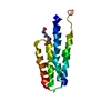
Mass: 307.323 Da / Num. of mol.: 1 / Source method: obtained synthetically / Formula: C10H17N3O6S

-
Experimental details

-
Experiment

ExperimentMethod: ELECTRON CRYSTALLOGRAPHY
EM experimentAggregation state: 2D ARRAY / 3D reconstruction method: electron crystallography

-
Sample preparation

ComponentName: Microsomal Glutathione Transferase 1 in Complex with Glutathione
Type: COMPLEX / Details: Embedded in trehalose
Buffer solutionName: potassium phosphate / pH: 8 / Details: potassium phosphate
SpecimenConc.: 1 mg/ml / Embedding applied: NO / Shadowing applied: NO / Staining applied: NO / Vitrification applied: YES
Specimen supportDetails: Carbon coated Mo-grids

-
Data collection

MicroscopyModel: JEOL 3000SFF / Details: Electron diffraction at 1.2 m camera length
Electron gunElectron source: FIELD EMISSION GUN / Accelerating voltage: 300 kV / Illumination mode: FLOOD BEAM
Electron lensMode: BRIGHT FIELD / Nominal magnification: 60000 X / Nominal defocus max: 2000 nm / Nominal defocus min: 300 nm / Cs: 1.6 mm
Specimen holderTemperature: 4 K / Tilt angle max: 62.6 ° / Tilt angle min: 0 °
Image recordingElectron dose: 10 e/Å2
Details: Kodak SO163 film for images, CCD for electron diffraction patterns
RadiationProtocol: SINGLE WAVELENGTH / Monochromatic (M) / Laue (L): M / Scattering type: electron
Radiation wavelengthRelative weight: 1

-
Processing

SoftwareName: REFMAC / Version: 5.2.0005 / Classification: refinement
CTF correctionDetails: Crystallographic
3D reconstructionMethod: Crystallographic / Resolution: 3.2 Å / Nominal pixel size: 1.17 Å
Magnification calibration: Gold electron diffraction to establish exact unit cell parameters
Symmetry type: 2D CRYSTAL
RefinementResolution: 3.2→10 Å / Cor.coef. Fo:Fc: 0.619 / Cor.coef. Fo:Fc free: 0.356 / SU B: 29.134 / SU ML: 0.54 / Cross valid method: THROUGHOUT / ESU R: 1.747 / ESU R Free: 0.635 / Stereochemistry target values: MAXIMUM LIKELIHOOD / Details: HYDROGENS HAVE BEEN ADDED IN THE RIDING POSITIONS
RfactorNum. reflection% reflectionSelection details
Rfree0.37628 457 9.4 %RANDOM
Rwork0.33894 ---
obs0.34237 4409 79.25 %-
Solvent computationIon probe radii: 0.8 Å / Shrinkage radii: 0.8 Å / VDW probe radii: 1.2 Å / Solvent model: MASK
Displacement parametersBiso mean: 26.662 Å2
Baniso -1Baniso -2Baniso -3
1--2.58 Å2-1.29 Å20 Å2
2---2.58 Å20 Å2
3---3.88 Å2
Refinement stepCycle: LAST / Resolution: 3.2→10 Å
ProteinNucleic acidLigandSolventTotal
Num. atoms964 0 20 0 984
Refine LS restraints
Refine-IDTypeDev idealDev ideal targetNumber
ELECTRON CRYSTALLOGRAPHYr_bond_refined_d0.0130.0221004
ELECTRON CRYSTALLOGRAPHYr_bond_other_d
ELECTRON CRYSTALLOGRAPHYr_angle_refined_deg1.8631.991358
ELECTRON CRYSTALLOGRAPHYr_angle_other_deg
ELECTRON CRYSTALLOGRAPHYr_dihedral_angle_1_deg9.8555119
ELECTRON CRYSTALLOGRAPHYr_dihedral_angle_2_deg36.93621.2241
ELECTRON CRYSTALLOGRAPHYr_dihedral_angle_3_deg26.2315165
ELECTRON CRYSTALLOGRAPHYr_dihedral_angle_4_deg16.716159
ELECTRON CRYSTALLOGRAPHYr_chiral_restr0.120.2159
ELECTRON CRYSTALLOGRAPHYr_gen_planes_refined0.0060.02742
ELECTRON CRYSTALLOGRAPHYr_gen_planes_other
ELECTRON CRYSTALLOGRAPHYr_nbd_refined0.3630.2806
ELECTRON CRYSTALLOGRAPHYr_nbd_other
ELECTRON CRYSTALLOGRAPHYr_nbtor_refined0.3360.2672
ELECTRON CRYSTALLOGRAPHYr_nbtor_other
ELECTRON CRYSTALLOGRAPHYr_xyhbond_nbd_refined0.2670.291
ELECTRON CRYSTALLOGRAPHYr_xyhbond_nbd_other
ELECTRON CRYSTALLOGRAPHYr_metal_ion_refined
ELECTRON CRYSTALLOGRAPHYr_metal_ion_other
ELECTRON CRYSTALLOGRAPHYr_symmetry_vdw_refined0.4140.269
ELECTRON CRYSTALLOGRAPHYr_symmetry_vdw_other
ELECTRON CRYSTALLOGRAPHYr_symmetry_hbond_refined0.3750.26
ELECTRON CRYSTALLOGRAPHYr_symmetry_hbond_other
ELECTRON CRYSTALLOGRAPHYr_symmetry_metal_ion_refined
ELECTRON CRYSTALLOGRAPHYr_symmetry_metal_ion_other
ELECTRON CRYSTALLOGRAPHYr_mcbond_it0.5291.5610
ELECTRON CRYSTALLOGRAPHYr_mcbond_other
ELECTRON CRYSTALLOGRAPHYr_mcangle_it0.9812970
ELECTRON CRYSTALLOGRAPHYr_scbond_it1.1313431
ELECTRON CRYSTALLOGRAPHYr_scangle_it1.6664.5388
ELECTRON CRYSTALLOGRAPHYr_rigid_bond_restr
ELECTRON CRYSTALLOGRAPHYr_sphericity_free
ELECTRON CRYSTALLOGRAPHYr_sphericity_bonded
LS refinement shellResolution: 3.2→3.274 Å / Total num. of bins used: 20
RfactorNum. reflection% reflection
Rfree0.398 29 -
Rwork0.314 262 -
obs--64.52 %

+
About Yorodumi

-
News

-
Feb 9, 2022. New format data for meta-information of EMDB entries

New format data for meta-information of EMDB entries

  • Version 3 of the EMDB header file is now the official format.
  • The previous official version 1.9 will be removed from the archive.

Related info.:EMDB header

External links:wwPDB to switch to version 3 of the EMDB data model

-
Aug 12, 2020. Covid-19 info

Covid-19 info

URL: https://pdbjlvh1.pdbj.org/emnavi/covid19.php

New page: Covid-19 featured information page in EM Navigator.

Related info.:Covid-19 info / Mar 5, 2020. Novel coronavirus structure data

+
Mar 5, 2020. Novel coronavirus structure data

Novel coronavirus structure data

Related info.:Yorodumi Speices / Aug 12, 2020. Covid-19 info

External links:COVID-19 featured content - PDBj / Molecule of the Month (242):Coronavirus Proteases

+
Jan 31, 2019. EMDB accession codes are about to change! (news from PDBe EMDB page)

EMDB accession codes are about to change! (news from PDBe EMDB page)

  • The allocation of 4 digits for EMDB accession codes will soon come to an end. Whilst these codes will remain in use, new EMDB accession codes will include an additional digit and will expand incrementally as the available range of codes is exhausted. The current 4-digit format prefixed with “EMD-” (i.e. EMD-XXXX) will advance to a 5-digit format (i.e. EMD-XXXXX), and so on. It is currently estimated that the 4-digit codes will be depleted around Spring 2019, at which point the 5-digit format will come into force.
  • The EM Navigator/Yorodumi systems omit the EMD- prefix.

Related info.:Q: What is EMD? / ID/Accession-code notation in Yorodumi/EM Navigator

External links:EMDB Accession Codes are Changing Soon! / Contact to PDBj

+
Jul 12, 2017. Major update of PDB

Major update of PDB

  • wwPDB released updated PDB data conforming to the new PDBx/mmCIF dictionary.
  • This is a major update changing the version number from 4 to 5, and with Remediation, in which all the entries are updated.
  • In this update, many items about electron microscopy experimental information are reorganized (e.g. em_software).
  • Now, EM Navigator and Yorodumi are based on the updated data.

External links:wwPDB Remediation / Enriched Model Files Conforming to OneDep Data Standards Now Available in the PDB FTP Archive

-
Yorodumi

Thousand views of thousand structures

  • Yorodumi is a browser for structure data from EMDB, PDB, SASBDB, etc.
  • This page is also the successor to EM Navigator detail page, and also detail information page/front-end page for Omokage search.
  • The word "yorodu" (or yorozu) is an old Japanese word meaning "ten thousand". "mi" (miru) is to see.

Related info.:EMDB / PDB / SASBDB / Comparison of 3 databanks / Yorodumi Search / Aug 31, 2016. New EM Navigator & Yorodumi / Yorodumi Papers / Jmol/JSmol / Function and homology information / Changes in new EM Navigator and Yorodumi

Read more