 Movie
Movie Controller


+ Open data

- Basic information
| Entry | Database: PDB / ID: 3qe7 | ||||||
|---|---|---|---|---|---|---|---|
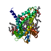
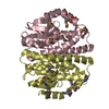
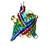
| Title | Crystal Structure of Uracil Transporter--UraA | ||||||
 Components Components | Uracil permease | ||||||
 Keywords Keywords | TRANSPORT PROTEIN / Uracil permease / Uracil transporter / UraA / transporter / inner membrane protein | ||||||
| Function / homology |  Function and homology information Function and homology informationuracil:monoatomic cation symporter activity / uracil transport / uracil import across plasma membrane / uracil transmembrane transporter activity / protein homodimerization activity / membrane / plasma membrane Similarity search - Function | ||||||
| Biological species |  | ||||||
| Method |  X-RAY DIFFRACTION / SYNCHROTRON / SAD / Resolution: 2.781 Å X-RAY DIFFRACTION / SYNCHROTRON / SAD / Resolution: 2.781 Å | ||||||
 Authors Authors | Lu, F.R. / Li, S. / Yan, N. | ||||||
 Citation Citation | Journal: Nature / Year: 2011 Title: Structure and mechanism of the uracil transporter UraA Authors: Lu, F. / Li, S. / Jiang, Y. / Jiang, J. / Fan, H. / Lu, G. / Deng, D. / Dang, S. / Zhang, X. / Wang, J. / Yan, N. | ||||||
| History |
|
- Structure visualization
| Structure viewer | Molecule: MolmilJmol/JSmol |
|---|
- Downloads & links
-Download
| PDBx/mmCIF format | 3qe7.cif.gz | 165.3 KB | Display | PDBx/mmCIF format |
|---|---|---|---|---|
| PDB format | pdb3qe7.ent.gz | 131.7 KB | Display | PDB format |
| PDBx/mmJSON format | 3qe7.json.gz | Tree view | PDBx/mmJSON format | |
| Others |  Other downloads Other downloads |
-Validation report
| Arichive directory | https://data.pdbj.org/pub/pdb/validation_reports/qe/3qe7ftp://data.pdbj.org/pub/pdb/validation_reports/qe/3qe7 | HTTPS FTP |
|---|
-Related structure data
| Similar structure data |
|---|


-Links
 PDBj
PDBj- Assembly
| Deposited unit | 
| ||||||||
|---|---|---|---|---|---|---|---|---|---|
| 1 |
| ||||||||
| 2 | 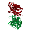
| ||||||||
| Unit cell |
|
-Components
| #1: Protein | Mass: 45089.805 Da / Num. of mol.: 1 Source method: isolated from a genetically manipulated source Source: (gene. exp.) |
|---|---|
| #2: Chemical | ChemComp-URA /   Mass: 112.087 Da / Num. of mol.: 1 / Source method: obtained synthetically / Formula: C4H4N2O2 Mass: 112.087 Da / Num. of mol.: 1 / Source method: obtained synthetically / Formula: C4H4N2O2 |
| #3: Sugar | ChemComp-BNG /   Type: D-saccharide / Mass: 306.395 Da / Num. of mol.: 1 Type: D-saccharide / Mass: 306.395 Da / Num. of mol.: 1Source method: isolated from a genetically manipulated source Formula: C15H30O6 / Comment: detergent*YM |
-Experimental details
-Experiment
| Experiment | Method: X-RAY DIFFRACTION / Number of used crystals: 1 |
|---|
- Sample preparation
| Crystal | Density Matthews: 3.78 Å3/Da / Density % sol: 67.42 % |
|---|---|
| Crystal grow | Temperature: 291 K / Method: vapor diffusion, hanging drop / pH: 6.5 Details: 25% PEG 400, 100mM MES-NaOH, 300mM Li2SO4, pH 6.5, VAPOR DIFFUSION, HANGING DROP, temperature 291K |
-Data collection
| Diffraction | Mean temperature: 100 K |
|---|---|
| Diffraction source | Source: SYNCHROTRON / Site: SPring-8  / Beamline: BL41XU / Wavelength: 0.99294 Å / Beamline: BL41XU / Wavelength: 0.99294 Å |
| Detector | Type: MARMOSAIC 225 mm CCD / Detector: CCD / Date: Apr 12, 2010 |
| Radiation | Monochromator: Mirror / Protocol: SINGLE WAVELENGTH / Monochromatic (M) / Laue (L): M / Scattering type: x-ray |
| Radiation wavelength | Wavelength: 0.99294 Å / Relative weight: 1 |
| Reflection | Resolution: 2.781→41.92 Å / Num. obs: 18225 / % possible obs: 98.7 % / Redundancy: 4.3 % / Biso Wilson estimate: 78.5 Å2 / Rmerge(I) obs: 0.078 / Net I/σ(I): 17.8 |
| Reflection shell | Resolution: 2.78→2.88 Å / Redundancy: 4.4 % / Rmerge(I) obs: 0.783 / Mean I/σ(I) obs: 1.9 / % possible all: 99.1 |
- Processing
| Software |
| ||||||||||||||||||||||||||||||||||||||||||||||||||||||||||||||||||||||||||||||||||||||||||||||||||||
|---|---|---|---|---|---|---|---|---|---|---|---|---|---|---|---|---|---|---|---|---|---|---|---|---|---|---|---|---|---|---|---|---|---|---|---|---|---|---|---|---|---|---|---|---|---|---|---|---|---|---|---|---|---|---|---|---|---|---|---|---|---|---|---|---|---|---|---|---|---|---|---|---|---|---|---|---|---|---|---|---|---|---|---|---|---|---|---|---|---|---|---|---|---|---|---|---|---|---|---|---|---|
| Refinement | Method to determine structure: SAD / Resolution: 2.781→41.92 Å / FOM work R set: 0.772 / SU ML: 0.46 / σ(F): 1.34 / Phase error: 28.13 / Stereochemistry target values: ML
| ||||||||||||||||||||||||||||||||||||||||||||||||||||||||||||||||||||||||||||||||||||||||||||||||||||
| Solvent computation | Shrinkage radii: 0.61 Å / VDW probe radii: 0.9 Å / Solvent model: FLAT BULK SOLVENT MODEL / Bsol: 69.203 Å2 / ksol: 0.303 e/Å3 | ||||||||||||||||||||||||||||||||||||||||||||||||||||||||||||||||||||||||||||||||||||||||||||||||||||
| Displacement parameters | Biso mean: 77.2341 Å2
| ||||||||||||||||||||||||||||||||||||||||||||||||||||||||||||||||||||||||||||||||||||||||||||||||||||
| Refinement step | Cycle: LAST / Resolution: 2.781→41.92 Å
| ||||||||||||||||||||||||||||||||||||||||||||||||||||||||||||||||||||||||||||||||||||||||||||||||||||
| Refine LS restraints |
| ||||||||||||||||||||||||||||||||||||||||||||||||||||||||||||||||||||||||||||||||||||||||||||||||||||
| LS refinement shell |
| ||||||||||||||||||||||||||||||||||||||||||||||||||||||||||||||||||||||||||||||||||||||||||||||||||||
| Refinement TLS params. | Method: refined / Refine-ID: X-RAY DIFFRACTION
| ||||||||||||||||||||||||||||||||||||||||||||||||||||||||||||||||||||||||||||||||||||||||||||||||||||
| Refinement TLS group |
|
