 Movie
Movie Controller

[English] 日本語
 Yorodumi
- PDB-3dww: Electron crystallographic structure of human microsomal prostagla... -
+ Open data

- Basic information
| Entry | Database: PDB / ID: 3dww | ||||||
|---|---|---|---|---|---|---|---|
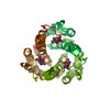
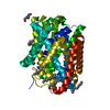
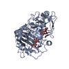
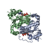
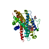
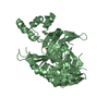
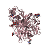
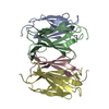
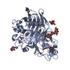
| Title | Electron crystallographic structure of human microsomal prostaglandin E synthase 1 | ||||||
 Components Components | Prostaglandin E synthase | ||||||
 Keywords Keywords | ISOMERASE / membrane protein / four helix bundle / Membrane / Transmembrane | ||||||
| Function / homology |  Function and homology information Function and homology informationprostaglandin-E synthase / prostaglandin-E synthase activity / regulation of fever generation / prostaglandin-D synthase activity / glutathione binding / cyclooxygenase pathway / positive regulation of prostaglandin secretion / Synthesis of Prostaglandins (PG) and Thromboxanes (TX) / glutathione peroxidase activity / prostaglandin biosynthetic process ...prostaglandin-E synthase / prostaglandin-E synthase activity / regulation of fever generation / prostaglandin-D synthase activity / glutathione binding / cyclooxygenase pathway / positive regulation of prostaglandin secretion / Synthesis of Prostaglandins (PG) and Thromboxanes (TX) / glutathione peroxidase activity / prostaglandin biosynthetic process / prostaglandin metabolic process / nuclear envelope lumen / glutathione transferase / glutathione transferase activity / Oxidoreductases; Acting on a peroxide as acceptor; Peroxidases / sensory perception of pain / regulation of inflammatory response / cell population proliferation / negative regulation of cell population proliferation / endoplasmic reticulum membrane / perinuclear region of cytoplasm / signal transduction / membrane Similarity search - Function | ||||||
| Biological species |  Homo sapiens (human) Homo sapiens (human) | ||||||
| Method | ELECTRON CRYSTALLOGRAPHY / electron crystallography / cryo EM / Resolution: 3.5 Å | ||||||
 Authors Authors | Hebert, H. / Jegerschold, C. | ||||||
 Citation Citation | Journal: Proc Natl Acad Sci U S A / Year: 2008 Title: Structural basis for induced formation of the inflammatory mediator prostaglandin E2. Authors: Caroline Jegerschöld / Sven-Christian Pawelzik / Pasi Purhonen / Priyaranjan Bhakat / Karina Roxana Gheorghe / Nobuhiko Gyobu / Kaoru Mitsuoka / Ralf Morgenstern / Per-Johan Jakobsson / Hans Hebert /  Abstract: Prostaglandins (PG) are bioactive lipids produced from arachidonic acid via the action of cyclooxygenases and terminal PG synthases. Microsomal prostaglandin E synthase 1 (MPGES1) constitutes an ...Prostaglandins (PG) are bioactive lipids produced from arachidonic acid via the action of cyclooxygenases and terminal PG synthases. Microsomal prostaglandin E synthase 1 (MPGES1) constitutes an inducible glutathione-dependent integral membrane protein that catalyzes the oxidoreduction of cyclooxygenase derived PGH(2) into PGE(2). MPGES1 has been implicated in a number of human diseases or pathological conditions, such as rheumatoid arthritis, fever, and pain, and is therefore regarded as a primary target for development of novel antiinflammatory drugs. To provide a structural basis for insight in the catalytic mechanism, we determined the structure of MPGES1 in complex with glutathione by electron crystallography from 2D crystals induced in the presence of phospholipids. Together with results from site-directed mutagenesis and activity measurements, we can thereby demonstrate the role of specific amino acid residues. Glutathione is found to bind in a U-shaped conformation at the interface between subunits in the protein trimer. It is exposed to a site facing the lipid bilayer, which forms the specific environment for the oxidoreduction of PGH(2) to PGE(2) after displacement of the cytoplasmic half of the N-terminal transmembrane helix. Hence, insight into the dynamic behavior of MPGES1 and homologous membrane proteins in inflammation and detoxification is provided. | ||||||
| History |
|
- Structure visualization
| Movie |
Movie viewer |
|---|---|
| Structure viewer | Molecule: MolmilJmol/JSmol |
- Downloads & links
-Download
| PDBx/mmCIF format | 3dww.cif.gz | 84.7 KB | Display | PDBx/mmCIF format |
|---|---|---|---|---|
| PDB format | pdb3dww.ent.gz | 64.3 KB | Display | PDB format |
| PDBx/mmJSON format | 3dww.json.gz | Tree view | PDBx/mmJSON format | |
| Others |  Other downloads Other downloads |
-Validation report
| Arichive directory | https://data.pdbj.org/pub/pdb/validation_reports/dw/3dwwftp://data.pdbj.org/pub/pdb/validation_reports/dw/3dww | HTTPS FTP |
|---|
-Related structure data
| Related structure data | |
|---|---|
| Similar structure data |
-Links
 PDBj
PDBj


- Assembly
| Deposited unit | 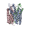
| ||||||||
|---|---|---|---|---|---|---|---|---|---|
| 1 |
| ||||||||
| Unit cell |
|
-Components
| #1: Protein | Mass: 17952.205 Da / Num. of mol.: 3 Source method: isolated from a genetically manipulated source Source: (gene. exp.) Homo sapiens (human) / Plasmid: pSP19T7LT / Production host:  #2: Chemical |   Mass: 307.323 Da / Num. of mol.: 3 / Source method: obtained synthetically / Formula: C10H17N3O6S Mass: 307.323 Da / Num. of mol.: 3 / Source method: obtained synthetically / Formula: C10H17N3O6S |
|---|
-Experimental details
-Experiment
| Experiment | Method: ELECTRON CRYSTALLOGRAPHY |
|---|---|
| EM experiment | Aggregation state: 2D ARRAY / 3D reconstruction method: electron crystallography |
- Sample preparation
| Component | Name: human microsomal prostaglandin E synthase 1 / Type: COMPLEX Details: Embedded in trehalose and frozen in liquid nitrogen |
|---|---|
| Specimen | Embedding applied: YES / Shadowing applied: NO / Staining applied: NO / Vitrification applied: YES |
| EM embedding | Details: 3 % trehalose / Material: trehalose |
-Data collection
| Microscopy | Model: JEOL 2100F / Details: Electron diffraction patterns |
|---|---|
| Electron gun | Electron source:  FIELD EMISSION GUN / Accelerating voltage: 200 kV / Illumination mode: FLOOD BEAM FIELD EMISSION GUN / Accelerating voltage: 200 kV / Illumination mode: FLOOD BEAM |
| Electron lens | Mode: DIFFRACTION |
| Specimen holder | Temperature: 100 K / Tilt angle max: 60 ° |
| Image recording | Film or detector model: TVIPS TEMCAM-F415 (4k x 4k) |
- Processing
| 3D reconstruction | Resolution: 3.5 Å Details: Phase information obtained by molecular replacement using the structure of microsomal glutathione transferase 1 Symmetry type: 2D CRYSTAL | ||||||||||||
|---|---|---|---|---|---|---|---|---|---|---|---|---|---|
| Refinement step | Cycle: LAST
|
