 Movie
Movie Controller

[English] 日本語
 Yorodumi
- PDB-2rg0: Crystal structure of cellobiohydrolase from Melanocarpus albomyce... -
+ Open data

- Basic information
| Entry | Database: PDB / ID: 2rg0 | ||||||||||||
|---|---|---|---|---|---|---|---|---|---|---|---|---|---|
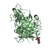
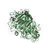
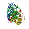
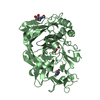
| Title | Crystal structure of cellobiohydrolase from Melanocarpus albomyces complexed with cellotetraose | ||||||||||||
 Components Components | Cellulose 1,4-beta-cellobiosidase | ||||||||||||
 Keywords Keywords | HYDROLASE / Glycosidase | ||||||||||||
| Function / homology |  Function and homology information Function and homology informationcellulose 1,4-beta-cellobiosidase activity / Hydrolases; Glycosylases; Glycosidases, i.e. enzymes that hydrolyse O- and S-glycosyl compounds / cellulose catabolic process Similarity search - Function | ||||||||||||
| Biological species |  Melanocarpus albomyces (fungus) Melanocarpus albomyces (fungus) | ||||||||||||
| Method |  X-RAY DIFFRACTION / SYNCHROTRON / MOLECULAR REPLACEMENT / Resolution: 2.1 Å X-RAY DIFFRACTION / SYNCHROTRON / MOLECULAR REPLACEMENT / Resolution: 2.1 Å | ||||||||||||
 Authors Authors | Parkkinen, T. / Koivula, A. / Vehmaanper, J. / Rouvinen, J. | ||||||||||||
 Citation Citation | Journal: Protein Sci. / Year: 2008 Title: Crystal structures of Melanocarpus albomyces cellobiohydrolase Cel7B in complex with cello-oligomers show high flexibility in the substrate binding Authors: Parkkinen, T. / Koivula, A. / Rouvinen, J. | ||||||||||||
| History |
|
- Structure visualization
| Structure viewer | Molecule: MolmilJmol/JSmol |
|---|
- Downloads & links
-Download
| PDBx/mmCIF format | 2rg0.cif.gz | 350.6 KB | Display | PDBx/mmCIF format |
|---|---|---|---|---|
| PDB format | pdb2rg0.ent.gz | 284.5 KB | Display | PDB format |
| PDBx/mmJSON format | 2rg0.json.gz | Tree view | PDBx/mmJSON format | |
| Others |  Other downloads Other downloads |
-Validation report
| Arichive directory | https://data.pdbj.org/pub/pdb/validation_reports/rg/2rg0ftp://data.pdbj.org/pub/pdb/validation_reports/rg/2rg0 | HTTPS FTP |
|---|
-Related structure data


-Links
 PDBj
PDBj- Assembly
| Deposited unit | 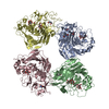
| ||||||||
|---|---|---|---|---|---|---|---|---|---|
| 1 | 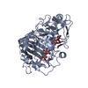
| ||||||||
| 2 | 
| ||||||||
| 3 | 
| ||||||||
| 4 | 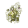
| ||||||||
| Unit cell |
|
-Components
| #1: Protein | Mass: 47572.230 Da / Num. of mol.: 4 / Mutation: Q1(PCA) Source method: isolated from a genetically manipulated source Source: (gene. exp.) Melanocarpus albomyces (fungus) / Production host: Trichoderma reesei (fungus)References: UniProt: Q8J0K6, cellulose 1,4-beta-cellobiosidase (non-reducing end) #2: Polysaccharide | beta-D-glucopyranose-(1-4)-beta-D-glucopyranose / beta-cellobiose   Source method: isolated from a genetically manipulated source Details: oligosaccharide / References: beta-cellobiose #3: Polysaccharide | beta-D-glucopyranose-(1-4)-beta-D-glucopyranose-(1-4)-beta-D-glucopyranose-(1-4)-beta-D-glucopyranose / beta-cellotetraose | 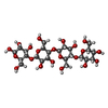  Source method: isolated from a genetically manipulated source Details: oligosaccharide / References: beta-cellotetraose #4: Water | ChemComp-HOH / |  Mass: 18.015 Da / Num. of mol.: 464 / Source method: isolated from a natural source / Formula: H2O Mass: 18.015 Da / Num. of mol.: 464 / Source method: isolated from a natural source / Formula: H2OHas protein modification | Y | |
|---|
-Experimental details
-Experiment
| Experiment | Method: X-RAY DIFFRACTION / Number of used crystals: 1 |
|---|
- Sample preparation
| Crystal | Density Matthews: 2.42 Å3/Da / Density % sol: 49.14 % |
|---|---|
| Crystal grow | Temperature: 298 K / Method: vapor diffusion, hanging drop / pH: 6.5 Details: 12% PEG8000, 0.1M calcium chloride, 0.1M cacodylate, pH6.5, VAPOR DIFFUSION, HANGING DROP, temperature 298K |
-Data collection
| Diffraction | Mean temperature: 100 K |
|---|---|
| Diffraction source | Source: SYNCHROTRON / Site: EMBL/DESY, HAMBURG  / Beamline: X11 / Wavelength: 0.8162 Å / Beamline: X11 / Wavelength: 0.8162 Å |
| Detector | Type: MAR CCD 165 mm / Detector: CCD / Date: Oct 3, 2006 |
| Radiation | Monochromator: Si(111) / Protocol: SINGLE WAVELENGTH / Monochromatic (M) / Laue (L): M / Scattering type: x-ray |
| Radiation wavelength | Wavelength: 0.8162 Å / Relative weight: 1 |
| Reflection | Resolution: 2.1→25 Å / Num. all: 105275 / Num. obs: 105275 / % possible obs: 99.5 % / Redundancy: 3.8 % / Biso Wilson estimate: 17.8 Å2 / Rsym value: 0.177 / Net I/σ(I): 7.6 |
| Reflection shell | Resolution: 2.1→2.2 Å / Mean I/σ(I) obs: 3.1 / Rsym value: 0.453 / % possible all: 97.9 |
- Processing
| Software |
| |||||||||||||||||||||||||||||||||
|---|---|---|---|---|---|---|---|---|---|---|---|---|---|---|---|---|---|---|---|---|---|---|---|---|---|---|---|---|---|---|---|---|---|---|
| Refinement | Method to determine structure: MOLECULAR REPLACEMENT / Resolution: 2.1→20 Å / Num. parameters: 55836 / Num. restraintsaints: 57366 / Cross valid method: FREE R / σ(F): 0 / Stereochemistry target values: ENGH AND HUBER / Details: Used twin operator h, -k, -l
| |||||||||||||||||||||||||||||||||
| Solvent computation | Solvent model: MOEWS & KRETSINGER | |||||||||||||||||||||||||||||||||
| Displacement parameters | Biso mean: 32.843 Å2 | |||||||||||||||||||||||||||||||||
| Refinement step | Cycle: LAST / Resolution: 2.1→20 Å
| |||||||||||||||||||||||||||||||||
| Refine LS restraints |
|
