 Movie
Movie Controller


+ Open data

- Basic information
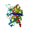
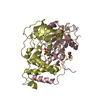
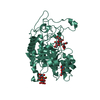
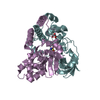
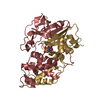
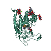
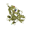
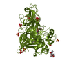
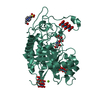
| Entry | Database: PDB / ID: 3nvj | ||||||
|---|---|---|---|---|---|---|---|
| Title | Crystal structure of the C143A/C166A mutant of Ero1p | ||||||
 Components Components | Endoplasmic oxidoreductin-1 | ||||||
 Keywords Keywords | OXIDOREDUCTASE / flavoenzyme / FAD / disulfide bonds / ER | ||||||
| Function / homology |  Function and homology information Function and homology informationthiol oxidase activity / thiol oxidase / protein folding in endoplasmic reticulum / protein-disulfide reductase activity / FAD binding / endoplasmic reticulum membrane / endoplasmic reticulum Similarity search - Function | ||||||
| Biological species |  | ||||||
| Method |  X-RAY DIFFRACTION / MOLECULAR REPLACEMENT / Resolution: 3.2 Å X-RAY DIFFRACTION / MOLECULAR REPLACEMENT / Resolution: 3.2 Å | ||||||
 Authors Authors | Fass, D. / Vonshak, O. | ||||||
 Citation Citation | Journal: Protein Sci. / Year: 2010 Title: Steps in reductive activation of the disulfide-generating enzyme Ero1p Authors: Heldman, N. / Vonshak, O. / Sevier, C.S. / Vitu, E. / Mehlman, T. / Fass, D. | ||||||
| History |
|
- Structure visualization
| Structure viewer | Molecule: MolmilJmol/JSmol |
|---|
- Downloads & links
-Download
| PDBx/mmCIF format | 3nvj.cif.gz | 87.4 KB | Display | PDBx/mmCIF format |
|---|---|---|---|---|
| PDB format | pdb3nvj.ent.gz | 64.5 KB | Display | PDB format |
| PDBx/mmJSON format | 3nvj.json.gz | Tree view | PDBx/mmJSON format | |
| Others |  Other downloads Other downloads |
-Validation report
| Arichive directory | https://data.pdbj.org/pub/pdb/validation_reports/nv/3nvjftp://data.pdbj.org/pub/pdb/validation_reports/nv/3nvj | HTTPS FTP |
|---|
-Related structure data
| Related structure data | 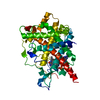 3m31C 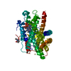 1rq1S S: Starting model for refinement C: citing same article ( |
|---|---|
| Similar structure data |


-Links
 PDBj
PDBj- Assembly
| Deposited unit | 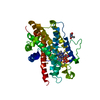
| ||||||||
|---|---|---|---|---|---|---|---|---|---|
| 1 |
| ||||||||
| Unit cell |
|
-Components
| #1: Protein | Mass: 45294.898 Da / Num. of mol.: 1 / Fragment: residues in UNP 56-424 / Mutation: C143A,C166A Source method: isolated from a genetically manipulated source Source: (gene. exp.) Gene: ERO1, YML130C, YM4987.05C / Plasmid: pGEX-4T1 / Production host:  References: UniProt: Q03103, Oxidoreductases; Acting on a sulfur group of donors; With a disulfide as acceptor |
|---|---|
| #2: Chemical | ChemComp-NEN /   Mass: 127.141 Da / Num. of mol.: 1 / Source method: obtained synthetically / Formula: C6H9NO2 Mass: 127.141 Da / Num. of mol.: 1 / Source method: obtained synthetically / Formula: C6H9NO2 |
| #3: Chemical | ChemComp-FAD /   Mass: 785.550 Da / Num. of mol.: 1 / Source method: obtained synthetically / Formula: C27H33N9O15P2 / Comment: FAD*YM Mass: 785.550 Da / Num. of mol.: 1 / Source method: obtained synthetically / Formula: C27H33N9O15P2 / Comment: FAD*YM |
| #4: Chemical | ChemComp-CD /   Mass: 112.411 Da / Num. of mol.: 1 / Source method: obtained synthetically / Formula: Cd Mass: 112.411 Da / Num. of mol.: 1 / Source method: obtained synthetically / Formula: Cd |
| #5: Water | ChemComp-HOH /  Mass: 18.015 Da / Num. of mol.: 12 / Source method: isolated from a natural source / Formula: H2O Mass: 18.015 Da / Num. of mol.: 12 / Source method: isolated from a natural source / Formula: H2O |
| Has protein modification | Y |
-Experimental details
-Experiment
| Experiment | Method: X-RAY DIFFRACTION / Number of used crystals: 1 |
|---|
- Sample preparation
| Crystal | Density Matthews: 4.64 Å3/Da / Density % sol: 73.51 % |
|---|
-Data collection
| Diffraction source | Source: ROTATING ANODE / Type: RIGAKU / Wavelength: 1.514 Å |
|---|---|
| Detector | Type: RIGAKU RAXIS IV++ / Detector: IMAGE PLATE / Date: May 16, 2010 |
| Radiation | Protocol: SINGLE WAVELENGTH / Monochromatic (M) / Laue (L): M / Scattering type: x-ray |
| Radiation wavelength | Wavelength: 1.514 Å / Relative weight: 1 |
| Reflection | Resolution: 3.2→50 Å / Num. all: 13439 / Num. obs: 13131 / % possible obs: 97.7 % / Observed criterion σ(F): 2 / Observed criterion σ(I): 1 |
- Processing
| Software |
| |||||||||||||||
|---|---|---|---|---|---|---|---|---|---|---|---|---|---|---|---|---|
| Refinement | Method to determine structure: MOLECULAR REPLACEMENT Starting model: 1RQ1 Resolution: 3.2→50 Å / σ(F): 2
| |||||||||||||||
| Refinement step | Cycle: LAST / Resolution: 3.2→50 Å
|
