 Movie
Movie Controller

[English] 日本語
 Yorodumi
- PDB-2h6r: Crystal Structure of triosephosphate isomerase (TIM) from Methano... -
+ Open data

- Basic information
| Entry | Database: PDB / ID: 2h6r | ||||||
|---|---|---|---|---|---|---|---|
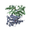
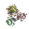
| Title | Crystal Structure of triosephosphate isomerase (TIM) from Methanocaldococcus jannaschii | ||||||
 Components Components | Triosephosphate isomerase | ||||||
 Keywords Keywords | ISOMERASE / beta-alpha barrel | ||||||
| Function / homology |  Function and homology information Function and homology informationtriose-phosphate isomerase / triose-phosphate isomerase activity / glyceraldehyde-3-phosphate biosynthetic process / glycerol catabolic process / gluconeogenesis / glycolytic process / cytosol Similarity search - Function | ||||||
| Biological species |   Methanocaldococcus jannaschii (archaea) Methanocaldococcus jannaschii (archaea) | ||||||
| Method |  X-RAY DIFFRACTION / SYNCHROTRON / MOLECULAR REPLACEMENT / Resolution: 2.3 Å X-RAY DIFFRACTION / SYNCHROTRON / MOLECULAR REPLACEMENT / Resolution: 2.3 Å | ||||||
 Authors Authors | Gayathri, P. / Banerjee, M. / Vijayalakshmi, A. / Balaram, H. / Balaram, P. / Murthy, M.R.N. | ||||||
 Citation Citation | Journal: Acta Crystallogr.,Sect.D / Year: 2007 Title: Structure of triosephosphate isomerase (TIM) from Methanocaldococcus jannaschii Authors: Gayathri, P. / Banerjee, M. / Vijayalakshmi, A. / Azeez, S. / Balaram, H. / Balaram, P. / Murthy, M.R.N. | ||||||
| History |
|
- Structure visualization
| Structure viewer | Molecule: MolmilJmol/JSmol |
|---|
- Downloads & links
-Download
| PDBx/mmCIF format | 2h6r.cif.gz | 305.6 KB | Display | PDBx/mmCIF format |
|---|---|---|---|---|
| PDB format | pdb2h6r.ent.gz | 248.8 KB | Display | PDB format |
| PDBx/mmJSON format | 2h6r.json.gz | Tree view | PDBx/mmJSON format | |
| Others |  Other downloads Other downloads |
-Validation report
| Arichive directory | https://data.pdbj.org/pub/pdb/validation_reports/h6/2h6rftp://data.pdbj.org/pub/pdb/validation_reports/h6/2h6r | HTTPS FTP |
|---|
-Related structure data
| Related structure data | 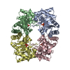 1hg3S S: Starting model for refinement |
|---|---|
| Similar structure data |


-Links
 PDBj
PDBj
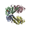
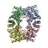
- Assembly
| Deposited unit | 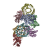
| ||||||||
|---|---|---|---|---|---|---|---|---|---|
| 1 | 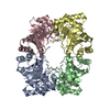
| ||||||||
| 2 | 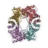
| ||||||||
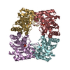
| Unit cell |
|
-Components
| #1: Protein | Mass: 23271.996 Da / Num. of mol.: 8 Source method: isolated from a genetically manipulated source Source: (gene. exp.) Methanocaldococcus jannaschii (archaea)Strain: DSM 2661 / Gene: TPIA / Plasmid: pTrc99A / Production host:  #2: Water | ChemComp-HOH / |  Mass: 18.015 Da / Num. of mol.: 384 / Source method: isolated from a natural source / Formula: H2O Mass: 18.015 Da / Num. of mol.: 384 / Source method: isolated from a natural source / Formula: H2O |
|---|
-Experimental details
-Experiment
| Experiment | Method: X-RAY DIFFRACTION / Number of used crystals: 1 |
|---|
- Sample preparation
| Crystal | Density Matthews: 2.43 Å3/Da / Density % sol: 49.44 % |
|---|---|
| Crystal grow | Temperature: 291 K / Method: microbatch / pH: 8 Details: 30% PEG 3350, 0.2M Magnesium chloride, 0.1M Tris pH 8.0, microbatch, temperature 291K |
-Data collection
| Diffraction | Mean temperature: 100 K | ||||||||||||||||||||||||||||||
|---|---|---|---|---|---|---|---|---|---|---|---|---|---|---|---|---|---|---|---|---|---|---|---|---|---|---|---|---|---|---|---|
| Diffraction source | Source: SYNCHROTRON / Site: SPring-8  / Beamline: BL44XU / Wavelength: 0.9 Å / Beamline: BL44XU / Wavelength: 0.9 Å | ||||||||||||||||||||||||||||||
| Detector | Type: MAC Science DIP-2040 / Detector: IMAGE PLATE / Date: Mar 20, 2006 | ||||||||||||||||||||||||||||||
| Radiation | Protocol: SINGLE WAVELENGTH / Monochromatic (M) / Laue (L): M / Scattering type: x-ray | ||||||||||||||||||||||||||||||
| Radiation wavelength | Wavelength: 0.9 Å / Relative weight: 1 | ||||||||||||||||||||||||||||||
| Reflection twin |
| ||||||||||||||||||||||||||||||
| Reflection | Resolution: 2.3→50 Å / Num. all: 76266 / Num. obs: 76266 / % possible obs: 96.3 % / Redundancy: 14.26 % / Biso Wilson estimate: 23.5 Å2 / Rmerge(I) obs: 0.094 / Net I/σ(I): 20.15 | ||||||||||||||||||||||||||||||
| Reflection shell | Resolution: 2.3→2.38 Å / Redundancy: 3.7 % / Rmerge(I) obs: 0.261 / Mean I/σ(I) obs: 6.3 / Num. unique all: 7537 |
- Processing
| Software |
| |||||||||||||||||||||||||
|---|---|---|---|---|---|---|---|---|---|---|---|---|---|---|---|---|---|---|---|---|---|---|---|---|---|---|
| Refinement | Method to determine structure: MOLECULAR REPLACEMENT Starting model: PDB entry 1HG3 Resolution: 2.3→34.28 Å / Rfactor Rfree error: 0.006 / Data cutoff high absF: 80028.33 / Data cutoff low absF: 0 / Isotropic thermal model: RESTRAINED / Cross valid method: THROUGHOUT / σ(F): 0 / Stereochemistry target values: Engh & Huber Details: The structure was refined using modified CNS files for tetartohedral twinning (Barends, et al., 2005, Acta Cryst D61, 613-621). The crystal structure has been determined from a ...Details: The structure was refined using modified CNS files for tetartohedral twinning (Barends, et al., 2005, Acta Cryst D61, 613-621). The crystal structure has been determined from a tetartohedrally twinned crystal. The twinning operator are (h,k,l)(k,h,-l)(-k,-h,-l)(-h,-k,l) and the twinning fraction are 0.25, 0.25, 0.25, 0.25.
| |||||||||||||||||||||||||
| Solvent computation | Solvent model: FLAT MODEL / Bsol: 49.1616 Å2 / ksol: 0.364509 e/Å3 | |||||||||||||||||||||||||
| Displacement parameters | Biso mean: 36.9 Å2
| |||||||||||||||||||||||||
| Refine analyze |
| |||||||||||||||||||||||||
| Refinement step | Cycle: LAST / Resolution: 2.3→34.28 Å
| |||||||||||||||||||||||||
| Refine LS restraints |
| |||||||||||||||||||||||||
| LS refinement shell | Resolution: 2.3→2.4 Å / Rfactor Rfree error: 0.018 / Total num. of bins used: 8
| |||||||||||||||||||||||||
| Xplor file |
|
